First Korean Case of Partial D DBS-1
- Affiliations
-
- 1Department of Laboratory Medicine, Soonchunhyang University Hospital Cheonan, Soonchunhyang University College of Medicine, Cheonan, Korea.
- 2Department of Health Sciences and Technology, Samsung Advanced Institute for Health Sciences and Technology, Sungkyunkwan University, Seoul, Korea. duck.cho@skku.edu
- 3Department of Laboratory Medicine and Genetics, Samsung Medical Center, Sungkyunkwan University School of Medicine, Seoul, Korea.
- KMID: 2470333
- DOI: http://doi.org/10.3343/alm.2020.40.4.337
Abstract
- No abstract available.
Figure
-
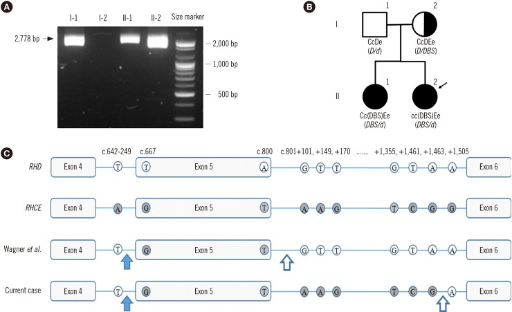
Fig. 1 Results of the genetic analysis of the proband and her family members. (A) Long-range PCR with primers located in non-Rhesus box sequences. A 2,778-bp fragment was amplified by PCR, indicating the presence of a hybrid RHD gene (lanes I-1, II-1, and II-2). (B) Pedigree, Rh phenotypes, and RHD genotypes of the Korean DBS-1 family. The genotypes and phenotypes of the DBS-1 family were determined using combined data from sequencing analysis, hybrid Rhesus box PCR, and serological analysis. Black circles indicate the DBS-1 phenotype. The proband is indicated by a black arrow. Total RHD deletion is denoted as “d” in the genotype. (C) Part of the RHD nucleotide sequence in DBS reported by Wagner, et al. [3] and this case. In both cases, the 5′ breakpoint region was located between the RHD-specific c.642-249T and the first RHCE-specific nucleotide, c.667G (blue arrow). The 3′ breakpoint region, located between the last RHCE-specific nucleotide and the first RHD-specific nucleotide of intron 5, differed for each case; it was located between c.800 and c.801+101 in the case reported by Wagner, et al. [3] and between c.801+1463 and c.801+1505 in the current case (white arrow).
Reference
-
1. Avent ND, Finning KM, Liu W, Scott ML. Molecular biology of partial D phenotypes. Transfus Clin Biol. 1996; 3:511–516. PMID: 9018818.2. Flegel WA, Von Zabern I, Doescher A, Wagner FF, Vytisková J, Písačka M. DCS-1, DCS-2, and DFV share amino acid substitutions at the extracellular RhD protein vestibule. Transfusion. 2008; 48:25–33. PMID: 17900276.3. Wagner FF, Ernst M, Sonneborn HH, Flegel WA. A DV-like phenotype is obliterated by A226P in the partial D DBS. Transfusion. 2001; 41:1052–1058. PMID: 11493738.4. Omi T, Takahashi J, Seno T, Tanaka M, Hirayama F, Matsuo M, et al. Isolation, characterization, and family study of DTI, a novel partial D phenotype affecting the fourth external loop of D polypeptides. Transfusion. 2002; 42:481–489. PMID: 12076297.5. Ye L, Wang P, Gao H, Zhang J, Wang C, Li Q, et al. Partial D phenotypes and genotypes in the Chinese population. Transfusion. 2012; 52:241–246. PMID: 21790636.6. Fasano RM, Monaco A, Meier ER, Pary P, Lee-Stroka AH, Otridge J, et al. RH genotyping in a sickle cell disease patient contributing to hematopoietic stem cell transplantation donor selection and management. Blood. 2010; 116:2836–2838. PMID: 20644109.7. Wagner F. The Human RhesusBase, version 2.4. Updated on Dec 2018. http://www.rhesusbase.info.8. Omi T, Okuda H, Iwamoto S, Kajii E, Takahashi J, Tanaka M, et al. Detection of Rh23 in the partial D phenotype associated with the DVa category. Transfusion. 2000; 40:256–257. PMID: 10686014.9. Wheeler MM, Lannert KW, Huston H, Fletcher SN, Harris S, Teramura G, et al. Genomic characterization of the RH locus detects complex and novel structural variation in multi-ethnic cohorts. Genet Med. 2019; 21:477. PMID: 29955105.10. Choi S, Chun S, Seo JY, Yang JH, Cho D. Planned transfusion of D-positive blood components in an Asia type DEL patient: proposed modification of the Korean National Guidelines for Blood Transfusion. Ann Lab Med. 2019; 39:102–104. PMID: 30215239.
- Full Text Links
-
- Actions
-
Cited
- CITED
-
- Close
- Share
-
- Similar articles
-
- Inadequate Efficacy of Deep Brain Stimulation in a Patient with Parkinson's disease due to Partial Breakage of Electrode Lead
- Deep Brain Stimulation of the Subthalamic and Pedunculopontine Nucleus in a Patient with Parkinson's Disease
- Deep Brain Stimulation for the Treatment of Movement Disorders
- Diffuse Brain Swelling in Severely Head Injured Patients
- Theoretical considerations of deep brain stimulation programming
