Korean J Ophthalmol.
2020 Feb;34(1):90-91. 10.3341/kjo.2019.0078.
Pigmented Paravenous Retinochoroidal Atrophy
- Affiliations
-
- 1Department of Ophthalmology, Seoul National University Bundang Hospital, Seoul National University College of Medicine, Seongnam, Korea. namooj@snubh.org
- 2Department of Ophthalmology, Seoul National University Hospital, Seoul National University College of Medicine, Seoul, Korea.
- KMID: 2469292
- DOI: http://doi.org/10.3341/kjo.2019.0078
Abstract
- No abstract available.
MeSH Terms
Figure
-
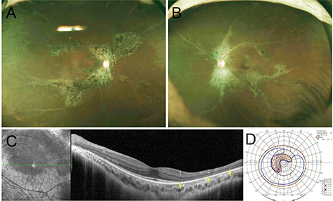
Fig. 1 The bony-spicule shaped retinochoroidal atrophy with pigmentation along retinal veins: wide fundus photography of the patient's (A) right eye and (B) left eye. (C) Optical coherence tomography of the patient's left eye. Diffuse disruption of the outer retina layer was observed while the microstructure of the macula remains intact. (D) Goldman perimetry examination of patient's left eye. Visual field is about 40 to 50 degrees; arcuate scotoma with enlarged blind spot corresponding to pigmented paravenous retinochoroidal atrophy presents.
Reference
-
1. Shen Y, Xu X, Cao H. Pigmented paravenous retinochoroidal atrophy: a case report. BMC Ophthalmol. 2018; 18:136.
Article2. Park HS, Yang JY, Park HJ. A case of unilateral focal pigmented paravenous retinochoroidal atrophy. J Korean Ophthalmol Soc. 2018; 59:1190–1194.
Article3. Choi JY, Sandberg MA, Berson EL. Natural course of ocular function in pigmented paravenous retinochoroidal atrophy. Am J Ophthalmol. 2006; 141:763–765.
Article4. Huang HB, Zhang YX. Pigmented paravenous retinochoroidal atrophy (review). Exp Ther Med. 2014; 7:1439–1445.
Article5. Traboulsi EI, Maumenee IH. Hereditary pigmented paravenous chorioretinal atrophy. Arch Ophthalmol. 1986; 104:1636–1640.
Article
- Full Text Links
-
- Actions
-
Cited
- CITED
-
- Close
- Share
-
- Similar articles
-
- Pigmented Paravenous Retinochoroidal Atrophy(PPRCA)
- A Case of Pigmented Paravenous Retino-Choroidal Atrophy and Retinitis Pigmentosa
- A Case of Unilateral Focal Pigmented Paravenous Retinochoroidal Atrophy
- A Case of Bultifocal choroiditis and Panuveitis
- Laser Treatment of a Retinochoroidal Coloboma Associated with Subreinal Neovascular Membrane
