Optimization of MRI Protocol for the Musculoskeletal System
- Affiliations
-
- 1Department of Radiology, Severance Hospital, Yonsei University College of Medicine, Seoul, Korea. radiologie@gmail.com
- 2Department of Radiology, Research Institute of Radiological Science, Center for Clinical Imaging Data Science (CCIDS), Yonsei University College of Medicine, Seoul, Korea.
- 3Department of Radiology, Gangnam Severance Hospital, Yonsei University College of Medicine, Seoul, Korea.
- KMID: 2469180
- DOI: http://doi.org/10.3348/jksr.2020.81.1.21
Abstract
- Magnetic resonance imaging (MRI) is an essential modality for the diagnosis of musculoskeletal system defects because of its higher soft-tissue contrast and spatial resolution. With the recent development of MRI-related technology, faster imaging and various image plane reconstructions are possible, enabling better assessment of three-dimensional musculoskeletal anatomy and lesions. Furthermore, the image quality, diagnostic accuracy, and acquisition time depend on the MRI protocol used. Moreover, the protocol affects the efficiency of the MRI scanner. Therefore, it is important for a radiologist to optimize the MRI protocol. In this review, we will provide guidance on patient positioning; selection of the radiofrequency coil, pulse sequences, and imaging planes; and control of MRI parameters to help optimize the MRI protocol for the six major joints of the musculoskeletal system.
MeSH Terms
Figure
-

Fig. 1. Dedicated shoulder RF coil. The shoulder is slightly externally rotated, and the shoulder cup of the RF coil is attached to the shoulder using the height adjustment bar. Since the shoulder joint is an off-center joint, it should be positioned at the center of the bore and the RF coil should be closely attached to the shoulder joint. RF = radiofrequency
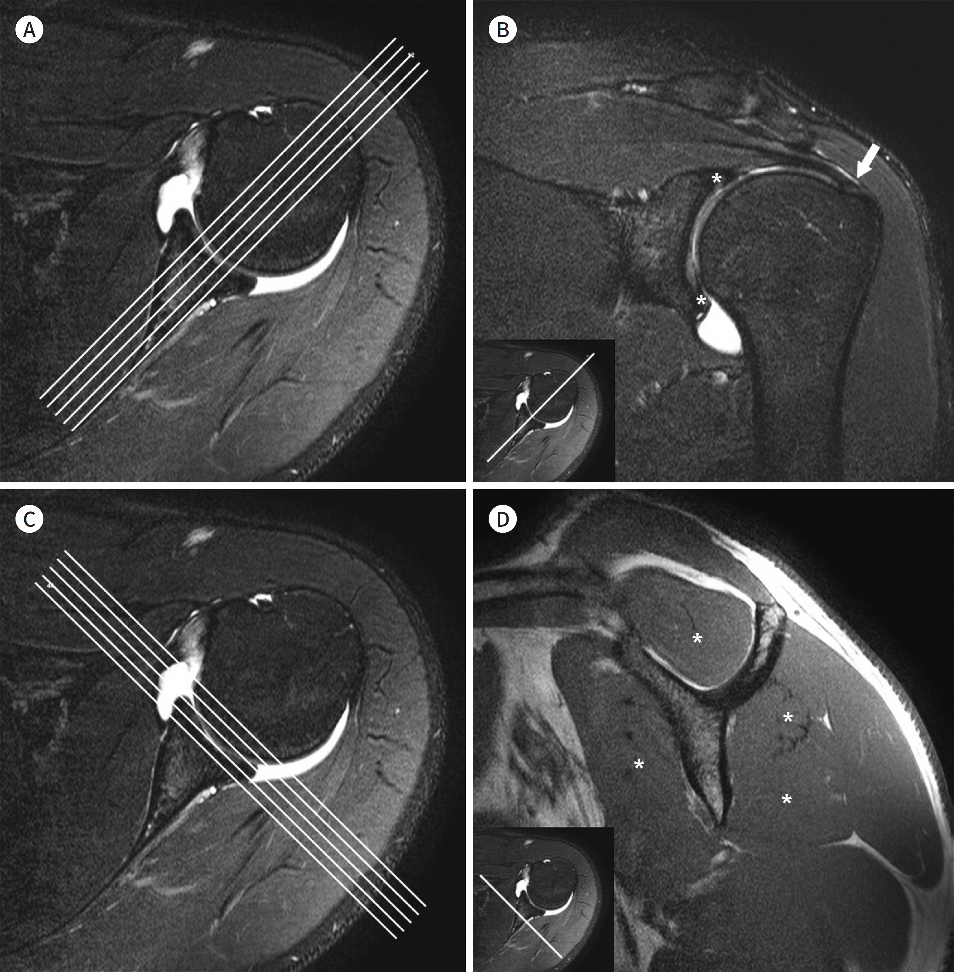
Fig. 2. Imaging plane of shoulder MRI. A-D. In shoulder MRI, based on axial images (A and C), the oblique coronal images (B) are obtained at right angles to the glenoid fossa (A), and the oblique sagittal images (D) are obtained perpendicular to the oblique coronal plane or parallel to the glenoid fossa (C). On the oblique coronal image (B), the glenoid labrum (asterisks) and the long course of the supraspinatus tendon and its attachment (arrow) to the greater tuberosity are well visualized. On the oblique sagittal image (D), the muscles forming the rotator cuff (asterisks) are visible. Clockwise from the 12 o’ clock position, the supraspinatus muscle, infraspinatus muscle, teres minor muscle, and subscapularis muscle are depicted.

Fig. 3. Elbow flexible RF coil. The coil is a flexible RF coil that can be used for small joints. The patient is in close contact with the elbow joint. Depending on the physique of the patient, small and medium sizes are available. RF = radiofrequency

Fig. 4. Dedicated wrist RF coil. By using the RF coil dedicated to the wrist joint, the patient can main-tain the joint in a neutral position while laying supine, thereby enabling stable image acquisition. The superman position may be considered to place the wrist joint at the center of the magnetic field. However, if the acquisition time is long, the patient may feel uncomfortable. RF = radiofrequency

Fig. 5. Torso RF coil for hip joint imaging. The coil is a multi-channel torso RF coil that can cover a wide field of view required for hip joint MRI, enabling imaging of both the hips and pelvic bones. RF = radiofrequency

Fig. 6. Dedicated knee RF coil. A type of dedicated RF coil for the knee joint, which can be separated up and down. When the knee joint is placed inside the coil, it is slightly flexed. Recently, radiologists have begun using the transmit-receiver coil, in which case the connectors are located on both sides. RF = radiofrequency

Fig. 7. Dedicated ankle RF coil. The ankle joint RF coil is designed so that the lower leg and the foot are al-most at right angles to each other, enabling correct posture for imaging. If a flexible coil is used, a positioner may be used to enhance patient comfort. RF = radiofrequency
Reference
-
1. Del Grande F, Santini F, Herzka DA, Aro MR, Dean CW, Gold GE, et al. Fat-suppression techniques for 3-T MR imaging of the musculoskeletal system.Radiographics. 2014; 34:217–233.2. Korean Society of Magnetic Resonance in Medicine. Clinical magnetic resonance imaging. Seoul: Ilchokak;2015.3. Jung JY, Yoon YC, Kwon JW, Ahn JH, Choe BK. Diagnosis of internal derangement of the knee at 3.0-T MR imaging: 3D isotropic intermediate-weighted versus 2D sequences.Radiology. 2009; 253:780–787.4. Hargreaves BA, Worters PW, Pauly KB, Pauly JM, Koch KM, Gold GE. Metal-induced artifacts in MRI. AJR Am J Roentgenol. 2011; 197:547–555.
Article5. Koch KM, Brau AC, Chen W, Gold GE, Hargreaves BA, Koff M, et al. Imaging near metal with a MAVRIC-SEMAC hybrid.Magn Reson Med. 2011; 65:71–82.6. American College of Radiology. ACR-SPR-SSR Practice parameter for the performance and interpretation of magnetic resonance imaging (MRI) of bone, joint, and soft tissue infections in the extremities. Avaiable at.https://www.acr.org/-/media/ACR/Files/Practice-Parameters/MR-Bone-Joint-Infections.pdf?la=en. Published 2016. Accessed Jan 10,. 2020.7. American College of Radiology. ACR-SPR-SSR practice parameter for the performance and interpretation of magnetic resonance imaging (MRI) of bone and soft tissue tumors. Avaiable at.https://www.acr.org/-/media/ACR/Files/Practice-Parameters/MR-SoftTissue-Tumors.pdf?la=en. Published 2015. Accessed Jan 10,. 2020.8. Steinbach LS, Palmer WE, Schweitzer ME. Special focus session: MR arthrography. Radiographics. 2002; 22:1223–1246.9. Chandnani VP, Yeager TD, DeBerardino T, Christensen K, Gagliardi JA, Heitz DR, et al. Glenoid labral tears: prospective evaluation with MRI imaging, MR arthrography, and CT arthrography.AJR Am J Roentgenol. 1993; 161:1229–1235.10. Cvitanic O, Tirman PF, Feller JF, Bost FW, Minter J, Carroll KW. Using abduction and external rotation of the shoulder to increase the sensitivity of MR arthrography in revealing tears of the anterior glenoid labrum.AJR Am J Roentgenol. 1997; 169:837–844.11. Davis SJ, Teresi LM, Bradley WG, Ressler JA, Eto RT. Effect of arm rotation on MR imaging of the rotator cuff. Radiology. 1991; 181:265–268.
Article12. Tuite MJ, De Smet AA, Norris MA, Orwin JF. MR diagnosis of labral tears of the shoulder: value of T2∗-weight-ed gradient-recalled echo images made in external rotation.AJR Am J Roentgenol. 1995; 164:941–944.13. Willemsen UF, Wiedemann E, Brunner U, Scheck R, Pfluger T, Kueffer G, et al. Prospective evaluation of MR arthrography performed with high-volume intraarticular saline enhancement in patients with recurrent anterior dislocations of the shoulder. AJR Am J Roentgenol. 1998; 170:79–84.
Article14. Jee WH. Musculoskeletal magnetic resonance imaging (MRI). Seoul: Korean Society of Musculoskeletal Radiology, Korean Society of Radiology;2017.15. Korean Society of Radiology. Quality assurance guidelines for special medical equipments. Seoul: Korean Institute for Accreditation of Medical Imaging;2019.16. Waldt S, Burkart A, Lange P, Imhoff AB, Rummeny EJ, Woertler K. Diagnostic performance of MR arthrography in the assessment of superior labral anteroposterior lesions of the shoulder.AJR Am J Roentgenol. 2004; 182:1271–1278.17. Palmer WE, Caslowitz PL. Anterior shoulder instability: diagnostic criteria determined from prospective analysis of 121 MR arthrograms. Radiology. 1995; 197:819–825.
Article18. Duc SR, Mengiardi B, Pfirrmann CW, Jost B, Hodler J, Zanetti M. Diagnostic performance of MR arthrography after rotator cuff repair. AJR Am J Roentgenol. 2006; 186:237–241.
Article19. Pfirrmann CW, Zanetti M, Weishaupt D, Gerber C, Hodler J. Subscapularis tendon tears: detection and grading at MR arthrography.Radiology. 1999; 213:709–714.20. Flannigan B, Kursunoglu-Brahme S, Snyder S, Karzel R, Del Pizzo W, Resnick D. MR arthrography of the shoulder: comparison with conventional MR imaging.AJR Am J Roentgenol. 1990; 155:829–832.21. Herold T, Bachthaler M, Hamer OW, Hente R, Feuerbach S, Fellner C, et al. Indirect MR arthrography of the shoulder: use of abduction and external rotation to detect full- and partial-thickness tears of the supraspinatus tendon. Radiology. 2006; 240:152–160.
Article22. Hodler J, Kursunoglu-Brahme S, Snyder SJ, Cervilla V, Karzel RP, Schweitzer ME, et al. Rotator cuff disease: assessment with MR arthrography versus standard MR imaging in 36 patients with arthroscopic confirma-tion.Radiology. 1992; 182:431–436.23. Palmer WE, Brown JH, Rosenthal DI. Rotator cuff: evaluation with fat-suppressed MR arthrography.Radiology. 1993; 188:683–687.24. Dinauer PA, Flemming DJ, Murphy KP, Doukas WC. Diagnosis of superior labral lesions: comparison of non-contrast MRI with indirect MR arthrography in unexercised shoulders. Skeletal Radiol. 2007; 36:195–202.
Article25. Kijowski R, Tuite M, Sanford M. Magnetic resonance imaging of the elbow. Part I: normal anatomy, imaging technique, and osseous abnormalities. Skeletal Radiol. 2004; 33:685–697.
Article26. Holtz P, Erickson SJ, Holmquist K. MR imaging of the elbow: technical considerations. Semin Musculoskelet Radiol. 1998; 2:121–132.
Article27. Rubin DA, Kneeland JB. MR imaging of the musculoskeletal system: technical considerations for enhancing image quality and diagnostic yield. AJR Am J Roentgenol. 1994; 163:1155–1163.
Article28. Kaplan LJ, Potter HG. MR imaging of ligament injuries to the elbow.Magn Reson Imaging Clin N Am. 2004; 12:221–232. v-vi.29. Kneeland JB. MR imaging of the elbow. Technical considerations.Magn Reson Imaging Clin N Am. 1997; 5:439–442.
Article30. Sonin AH, Fitzgerald SW. MR imaging of sports injuries in the adult elbow: a tailored approach. AJR Am J Roentgenol. 1996; 167:325–331.
Article31. Hunter JC, Escobedo EM, Wilson AJ, Hanel DP, Zink-Brody GC, Mann FA. MR imaging of clinically suspected scaphoid fractures. AJR Am J Roentgenol. 1997; 168:1287–1293.
Article32. Weiss KL, Beltran J, Shamam OM, Stilla RF, Levey M. High-field MR surface-coil imaging of the hand and wrist. Part I. Normal anatomy.Radiology. 1986; 160:143–146.33. Kulkarni MV, Patton JA, Price RR. Technical considerations for the use of surface coils in MRI.AJR Am J Roentgenol. 1986; 147:373–378.34. Amrami KK, Felmlee JP. 3-Tesla imaging of the wrist and hand: techniques and applications.Semin Musculoskelet Radiol. 2008; 12:223–237.35. Saupe N. 3-tesla high-resolution MR imaging of the wrist.Semin Musculoskelet Radiol. 2009; 13:29–38.36. American College of Radiology. ACR-SPR-SSR practice parameter for the performance and interpretation of magnetic resonance imaging of elbow. Avaiable at.https://www.acr.org/-/media/ACR/Files/Practice-Pa-rameters/MR-Elbow.pdf?la=en. Published 2016. Accessed Jan 10,. 2020.37. Scheck RJ, Romagnolo A, Hierner R, Pfluger T, Wilhelm K, Hahn K. The carpal ligaments in MR arthrography of the wrist: correlation with standard MRI and wrist arthroscopy.J Magn Reson Imaging. 1999; 9:468–474.38. Scheck RJ, Kubitzek C, Hierner R, Szeimies U, Pfluger T, Wilhelm K, et al. The scapholunate interosseous ligament in MR arthrography of the wrist: correlation with non-enhanced MRI and wrist arthroscopy.Skeletal Radiol. 1997; 26:263–271.39. Haims AH, Schweitzer ME, Morrison WB, Deely D, Lange RC, Osterman AL, et al. Internal derangement of the wrist: indirect MR arthrography versus unenhanced MR imaging. Radiology. 2003; 227:701–707.
Article40. Theumann NH, Pfirrmann CW, Chung CB, Antonio GE, Trudell DJ, Resnick D. Pisotriquetral joint: assessment with MR imaging and MR arthrography.Radiology. 2002; 222:763–770.41. Schweitzer ME, Natale P, Winalski CS, Culp R. Indirect wrist MR arthrography: the effects of passive motion versus active exercise. Skeletal Radiol. 2000; 29:10–14.
Article42. Hayes CW, Balkissoon AA. Magnetic resonance imaging of the musculoskeletal system. II. The hip.Clin Orthop Relat Res. 1996; 322:297–309.43. Sankey RA, Turner J, Lee J, Healy J, Gibbons CE. The use of MRI to detect occult fractures of the proximal femur: a study of 102 consecutive cases over a ten-year period. J Bone Joint Surg Br. 2009; 91:1064–1068.44. Khanna AJ, Yoon TR, Mont MA, Hungerford DS, Bluemke DA. Femoral head osteonecrosis: detection and grading by using a rapid MR imaging protocol.Radiology. 2000; 217:188–192.45. Bollow M, Braun J, Hamm B, Eggens U, Schilling A, König H, et al. Early sacroiliitis in patients with spondylo-arthropathy: evaluation with dynamic gadolinium-enhanced MR imaging.Radiology. 1995; 194:529–536.46. Murphey MD, Wetzel LH, Bramble JM, Levine E, Simpson KM, Lindsley HB. Sacroiliitis: MR imaging findings. Radiology. 1991; 180:239–244.
Article47. Horii M, Kubo T, Hirasawa Y. Radial MRI of the hip with moderate osteoarthritis.J Bone Joint Surg Br. 2000; 82:364–368.48. Dudda M, Albers C, Mamisch TC, Werlen S, Beck M. Do normal radiographs exclude asphericity of the femoral head-neck junction?Clin Orthop Relat Res. 2009; 467:651–659.49. Omar IM, Zoga AC, Kavanagh EC, Koulouris G, Bergin D, Gopez AG, et al. Athletic pubalgia and “sports her-nia”: optimal MR imaging technique and findings. Radiographics. 2008; 28:1415–1438.
Article50. Takao M, Sugano N, Nishii T, Tanaka H, Masumoto J, Miki H, et al. Application of three-dimensional magnet-ic resonance image registration for monitoring hip joint diseases.Magn Reson Imaging. 2005; 23:665–670.51. Cvitanic O, Henzie G, Skezas N, Lyons J, Minter J. MRI diagnosis of tears of the hip abductor tendons (gluteus medius and gluteus minimus). AJR Am J Roentgenol. 2004; 182:137–143.
Article52. Shuman WP, Castagno AA, Baron RL, Richardson ML. MR imaging of avascular necrosis of the femoral head: value of small-field-of-view sagittal surface-coil images. AJR Am J Roentgenol. 1988; 150:1073–1078.
Article53. Czerny C, Hofmann S, Urban M, Tschauner C, Neuhold A, Pretterklieber M, et al. MR arthrography of the adult acetabular capsular-labral complex: correlation with surgery and anatomy. AJR Am J Roentgenol. 1999; 173:345–349.
Article54. Magee T, Shapiro M, Williams D. Usefulness of simultaneous acquisition of spatial harmonics technique for MRI of the knee.AJR Am J Roentgenol. 2004; 182:1411–1415.55. Gold GE, Busse RF, Beehler C, Han E, Brau AC, Beatty PJ, et al. Isotropic MRI of the knee with 3D fast spin-echo extended echo-train acquisition (XETA): initial experience. AJR Am J Roentgenol. 2007; 188:1287–1293.
Article56. Gold GE, Hargreaves BA, Vasanawala SS, Webb JD, Shimakawa AS, Brittain JH, et al. Articular cartilage of the knee: evaluation with fluctuating equilibrium MR imaging–initial experience in healthy volunteers. Radiology. 2006; 238:712–718.
Article57. Buckwalter KA, Pennes DR. Anterior cruciate ligament: oblique sagittal MR imaging.Radiology. 1990; 175:276–277.58. Yu JS, Salonen DC, Hodler J, Haghighi P, Trudell D, Resnick D. Posterolateral aspect of the knee: improved MR imaging with a coronal oblique technique.Radiology. 1996; 198:199–204.59. Ha TP, Li KC, Beaulieu CF, Bergman G, Ch'en IY, Eller DJ, et al. Anterior cruciate ligament injury: fast spin-echo MR imaging with arthroscopic correlation in 217 examinations. AJR Am J Roentgenol. 1998; 170:1215–1219.
Article60. Mink JH, Levy T, Crues JV 3rd. Tears of the anterior cruciate ligament and menisci of the knee: MR imaging evaluation.Radiology. 1988; 167:769–774.61. Ho YY, Stanley AJ, Hui JH, Wang SC. Postoperative evaluation of the knee after autologous chondrocyte im-plantation: what radiologists need to know.Radiographics. 2007; 27:207–220.62. Kramer J, Recht MP, Imhof H, Stiglbaüer R, Engel A. Postcontrast MR arthrography in assessment of cartilage lesions.J Comput Assist Tomogr. 1994; 18:218–224.63. Brossmann J, Preidler KW, Daenen B, Pedowitz RA, Andresen R, Clopton P, et al. Imaging of osseous and cartilaginous intraarticular bodies in the knee: comparison of MR imaging and MR arthrography with CT and CT arthrography in cadavers.Radiology. 1996; 200:509–517.64. McCauley TR. MR imaging evaluation of the postoperative knee.Radiology. 2005; 234:53–61.65. Vahlensieck M, Peterfy CG, Wischer T, Sommer T, Lang P, Schlippert U, et al. Indirect MR arthrography: optimization and clinical applications.Radiology. 1996; 200:249–254.66. Peh WC, Chan JH. Artifacts in musculoskeletal magnetic resonance imaging: identification and correction. Skeletal Radiol. 2001; 30:179–191.
Article67. Van Hecke PE, Marchal GJ, Baert AL. Use of shielding to prevent folding in MR imaging.Radiology. 1988; 167:557–558.68. Magee T, Williams D. 3.0-T MRI of meniscal tears. AJR Am J Roentgenol. 2006; 187:371–375.
Article69. Shellock FG. Magnetic resonance procedures: health effects and safety. Boca Raton: Crc Press. 2000.70. Raphael B, Haims AH, Wu JS, Katz LD, White LM, Lynch K. MRI comparison of periprosthetic structures around zirconium knee prostheses and cobalt chrome prostheses.AJR Am J Roentgenol. 2006; 186:1771–1777.71. Mesgarzadeh M, Schneck CD, Tehranzadeh J, Chandnani VP, Bonakdarpour A. Magnetic resonance imaging of ankle ligaments. Emphasis on anatomy and injuries to lateral collateral ligaments.Magn Reson Imaging Clin N Am. 1994; 2:39–58.
Article72. Tokuda O, Awaya H, Taguchi K, Matsunga N. Kinematic MRI of the normal ankle ligaments using a specially designed passive positioning device.Foot Ankle Int. 2006; 27:935–942.73. Kneeland JB. Technical considerations for magnetic resonance imaging of the ankle and foot.Magn Reson Imaging Clin N Am. 1994; 2:23–28.74. Oae K, Takao M, Naito K, Uchio Y, Kono T, Ishida J, et al. Injury of the tibiofibular syndesmosis: value of MR imaging for diagnosis. Radiology. 2003; 227:155–161.
Article75. Barr C, Bauer JS, Malfair D, Ma B, Henning TD, Steinbach L, et al. MR imaging of the ankle at 3 Tesla and 1.5 Tesla: protocol optimization and application to cartilage, ligament and tendon pathology in cadaver speci-mens. Eur Radiol. 2007; 17:1518–1528.
Article76. Schibany N, Ba-Ssalamah A, Marlovits S, Mlynarik V, Nöbauer-Huhmann IM, Striessnig G, et al. Impact of high field (3.0 T) magnetic resonance imaging on diagnosis of osteochondral defects in the ankle joint.Eur J Radiol. 2005; 55:283–288.77. Moshirfar A, Campbell JT, Khanna AJ, Byank RP, Bluemke DA, Wenz JF Sr. Magnetic resonance imaging of the ankle: techniques and spectrum of disease.J Bone Joint Surg Am. 2003; 85-A(Suppl 4):7–19.78. Ramnath RR. 3T MR imaging of the musculoskeletal system (part II): clinical applications.Magn Reson Imaging Clin N Am. 2006; 14:41–62.79. Erickson SJ, Cox IH, Hyde JS, Carrera GF, Strandt JA, Estkowski LD. Effect of tendon orientation on MR imaging signal intensity: a manifestation of the “magic angle” phenomenon.Radiology. 1991; 181:389–392.
- Full Text Links
-
- Actions
-
Cited
- CITED
-
- Close
- Share
-
- Similar articles
-
- Introduction to Knobology Focusing on B Mode and Doppler Setting in Musculoskeletal Ultrasound
- Ultrasound-guided Intervention in Musculoskeletal System
- Menopause and Musculoskeletal Pain
- Amyloid-Related Imaging Abnormalities in Anti-Amyloid Monoclonal Antibody Therapy for Alzheimer’s Disease: Expert Recommendation for Standard MRI Protocol
- Abbreviated Breast Magnetic Resonance Imaging: Background, Evidence From Studies, and Future Considerations
