Chest CT Features of Cystic Fibrosis in Korea: Comparison with Non-Cystic Fibrosis Diseases
- Affiliations
-
- 1Department of Radiology and Center for Imaging Science, Samsung Medical Center, Sungkyunkwan University School of Medicine, Seoul 06351, Korea. kyungs.lee@samsung.com
- 2Department of Radiology, Chung-Ang University Hospital, Chung-Ang University College of Medicine, Seoul 06973, Korea.
- 3Department of Radiology, Hanyang University Hospital, Hanyang University College of Medicine, Seoul 04763, Korea.
- KMID: 2468138
- DOI: http://doi.org/10.3348/kjr.2017.18.1.260
Abstract
OBJECTIVE
Cystic fibrosis (CF) is a rare congenital disease in Korea, and its clinical and imaging findings are unclear. The objective of our study was to describe the clinical and CT features of CF in Korea and compare its features with those of other diseases mimicking CF.
MATERIALS AND METHODS
From November 1994 to December 2014, a presumptive diagnosis of CF was made in 23 patients based on clinical or radiological examination. After the exclusion of 10 patients without diagnostic confirmation, 13 patients were included in the study. A diagnosis of CF was made with the CF gene study. CT findings were evaluated for the presence and distribution of parenchymal abnormalities including bronchiectasis, tree-in-bud (TIB) pattern, mucus plugging, consolidation, and mosaic attenuation.
RESULTS
Of the 13 patients, 7 (median age, 15 years) were confirmed as CF, 4 (median age, 19 years) had primary ciliary dyskinesia, 1 had bronchiectasis of unknown cause, and 1 had chronic asthma. CT of patients with CF showed bilateral bronchiectasis, TIB pattern, mosaic attenuation, and mucus plugging in all patients, with upper lung predominance (57%). In CT of the non-CF patients, bilateral bronchiectasis, TIB pattern, mosaic attenuation, and mucus plugging were also predominant features, with lower lung predominance (50%).
CONCLUSION
Korean patients with CF showed bilateral bronchiectasis, cellular bronchiolitis, mucus plugging, and mosaic attenuation, which overlapped with those of non-CF patients. CF gene study is recommended for the definitive diagnosis of CF in patients with these clinical and imaging features.
MeSH Terms
Figure
-

Fig. 1 Flowchart of study population. CF = cystic fibrosis, CFTR = cystic fibrosis transmembrane conductance regulator, EM = electromicroscopic, PCD = primary ciliary dyskinesia

Fig. 2 Cystic fibrosis in 15-year-old male patient who underwent lung transplantation. Lung window image of thin-section (2.5-mm-section thickness) CT scan obtained at levels of aortic arch shows extensive areas of bronchiectasis (arrows) and cellular bronchiolitis (arrowheads) in both lungs. Also note patchy areas (open arrows) of mosaic attenuation.
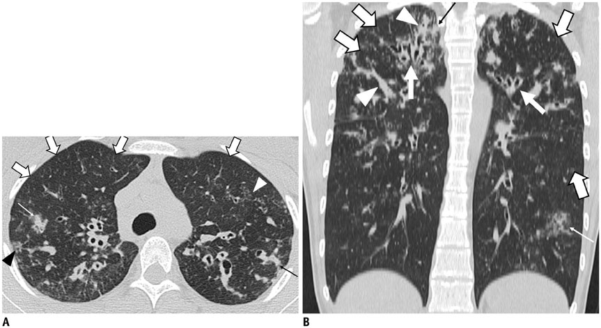
Fig. 3 17-year-old female patient with cystic fibrosis and Pseudomonas aeruginosa infection. A. Lung window image of thin-section (2.0-mm-section thickness) CT obtained at level of aortic arch shows extensive areas of bronchiectasis and cellular bronchiolitis (arrowheads) in both lungs. Also note wide areas (open arrows) of mosaic attenuation. Several areas of rectangular consolidation (thin arrows) suggest presence of concurrent bacterial pneumonia. B. Coronal reformatted (2.0-mm-section thickness) CT image demonstrates areas of bronchiectasis (arrows), mosaic attenuation (open arrows), and mucus plugging (arrowheads) predominantly involving bilateral upper lung zones. Also note areas of parenchymal opacity (thin arrows).

Fig. 4 19-year-old female patient with primary ciliary dyskinesia syndrome and concurrent infection with Mycobacterium gordonae. A. Lung window image of thin-section (2.5-mm-section thickness) CT obtained at level of lower esophagus shows extensive areas of bronchiectasis (arrows) and mucus plugging (arrowheads) in both lungs. Also note patchy areas (open arrows) of mosaic attenuation. B. Coronal reformatted (2.0-mm-section thickness) CT image demonstrates bronchiectasis (arrows) predominantly in lower lung zones. Also note mucus plugging (arrowhead) and parenchymal consolidation (thin arrows).
Cited by 1 articles
-
Age of Data in Contemporary Research Articles Published in Representative General Radiology Journals
Ji Hun Kang, Dong Hwan Kim, Seong Ho Park, Jung Hwan Baek
Korean J Radiol. 2018;19(6):1172-1178. doi: 10.3348/kjr.2018.19.6.1172.
Reference
-
1. Scriver CR, Beaudet AL, Sly WS, Valle D. The metabolic and molecular bases of inherited disease. 8th ed. New York, NY: McGraw-Hill;2001. p. 5121–5188.2. Yamashiro Y, Shimizu T, Oguchi S, Shioya T, Nagata S, Ohtsuka Y. The estimated incidence of cystic fibrosis in Japan. J Pediatr Gastroenterol Nutr. 1997; 24:544–547.3. Ahn KM, Park HY, Lee JH, Lee MG, Kim JH, Kang IJ, et al. Cystic fibrosis in Korean children: a case report identified by a quantitative pilocarpine iontophoresis sweat test and genetic analysis. J Korean Med Sci. 2005; 20:153–157.4. Choe YJ, Ko JS, Seo JK, Han JJ, Shim JO, Koh YY, et al. Novel CFTR mutations in a Korean infant with cystic fibrosis and pancreatic insufficiency. J Korean Med Sci. 2010; 25:163–165.5. Gee HY, Kim CK, Kim SW, Lee JH, Kim JH, Kim KH, et al. The L441P mutation of cystic fibrosis transmembrane conductance regulator and its molecular pathogenic mechanisms in a Korean patient with cystic fibrosis. J Korean Med Sci. 2010; 25:166–171.6. Hwang IO, Lee ES. A case of cystic fibrosis presented with meconium ileus in a female neonate. Korean J Pediatr. 2007; 50:1252–1256.7. Ko JM, Kim GH, Kim KM, Hong SJ, Yoo HW. Identification of a novel mutation of CFTR gene in a Korean patient with cystic fibrosis. J Korean Med Sci. 2008; 23:912–915.8. Koh WJ, Ki CS, Kim JW, Kim JH, Lim SY. Report of a Korean patient with cystic fibrosis, carrying Q98R and Q220X mutations in the CFTR gene. J Korean Med Sci. 2006; 21:563–566.9. Moon HR, Ko TS, Ko YY, Choi JH, Kim YC. Cystic fibrosis--a case presented with recurrent bronchiolitis in infancy in a Korean male infant. J Korean Med Sci. 1988; 3:157–162.10. Park SH, Lee Hj, Kim JH, Park CH. Cystic fibrosis: case report. J Korean Radiol Soc. 2002; 47:693–696.11. Pedrosa JF, da Cunha Ibiapina C, Alvim CG, Camargos PA, Martins FP, Guimarães EV, et al. Pulmonary radiographic findings in young children with cystic fibrosis. Pediatr Radiol. 2015; 45:153–157.12. Ernst CW, Basten IA, Ilsen B, Buls N, Van Gompel G, De Wachter E, et al. Pulmonary disease in cystic fibrosis: assessment with chest CT at chest radiography dose levels. Radiology. 2014; 273:597–605.13. Bhat V, Wahab AA, Garg KC, Janahi I, Singh R. HRCT in cystic fibrosis in patients with CFTR I1234V mutation: assessment of scoring systems with low dose technique using multidetector system and correlation with pulmonary function tests. Indian J Radiol Imaging. 2015; 25:44–51.14. Brody AS, Klein JS, Molina PL, Quan J, Bean JA, Wilmott RW. High-resolution computed tomography in young patients with cystic fibrosis: distribution of abnormalities and correlation with pulmonary function tests. J Pediatr. 2004; 145:32–38.15. Flight WG, Jones AM. Cystic fibrosis, primary ciliary dyskinesia and non-cystic fibrosis bronchiectasis: update 2008-11. Thorax. 2012; 67:645–649.16. Miller WT Jr, Panosian JS. Causes and imaging patterns of tree-in-bud opacities. Chest. 2013; 144:1883–1892.17. Hansell DM, Bankier AA, MacMahon H, McLoud TC, Müller NL, Remy J. Fleischner Society: glossary of terms for thoracic imaging. Radiology. 2008; 246:697–722.18. Jung H, Ki CS, Koh WJ, Ahn KM, Lee SI, Kim JH, et al. Heterogeneous spectrum of CFTR gene mutations in Korean patients with cystic fibrosis. Korean J Lab Med. 2011; 31:219–224.19. Kim SJ, Lee M, Cha SI, Park HY, Ahn KM, Ki CS, et al. [Standardized sweat chloride analysis for the diagnosis of cystic fibrosis in Korea]. Korean J Lab Med. 2008; 28:274–281.20. Kennedy MP, Noone PG, Leigh MW, Zariwala MA, Minnix SL, Knowles MR, et al. High-resolution CT of patients with primary ciliary dyskinesia. AJR Am J Roentgenol. 2007; 188:1232–1238.21. Rosenfeld M, Davis R, FitzSimmons S, Pepe M, Ramsey B. Gender gap in cystic fibrosis mortality. Am J Epidemiol. 1997; 145:794–803.22. Stephenson A, Hux J, Tullis E, Austin PC, Corey M, Ray J. Higher risk of hospitalization among females with cystic fibrosis. J Cyst Fibros. 2011; 10:93–99.23. Kim MJ, Kang JW, Lee JH, Kim KW, Sohn MH, Lee MG, et al. A case report of a classic cystic fibrosis pediatric patient in Korea carrying very rare CFTR gene mutations (D993Y and Q220X). Pediatr Allergy Respir Dis. 2011; 21:61–66.
- Full Text Links
-
- Actions
-
Cited
- CITED
-
- Close
- Share
-
- Similar articles
-
- Cystic Fibrosis: Case Report
- Cystic fibrosis lung disease: Current perspectives
- Clinical and Imaging Features of Cystic Fibrosis in Korean Children
- Cystic fibrosis in a female adolescent carrying c.263T>G (p.Leu88X) and c.2977G>T (p.Asp993Tyr) mutation
- A Case of Mycobacterium abscessus Lung Disease in a Patient with Cystic Fibrosis
