Predictors of the Response to an Epidural Blood Patch in Patients with Spinal Leakage of Cerebrospinal Fluid
- Affiliations
-
- 1Department of Neurosurgery, International University of Health and Welfare Atami Hospital, Atami, Shizuoka, Japan. hiroshikannomd@nifty.com, macshino@iuhw.ac.jp
- 2Department of Pharmacology, International University of Health and Welfare Atami Hospital, Atami, Shizuoka, Japan.
- KMID: 2467778
- DOI: http://doi.org/10.3988/jcn.2020.16.1.1
Abstract
- BACKGROUND AND PURPOSE
An epidural blood patch (EBP) is a highly effective therapy for spinal cerebrospinal fluid (CSF) leakage. However, the factors predicting the response to an EBP have not been fully elucidated. The aim of this study was to elucidate factors predicting the response to an EBP.
METHODS
We retrospectively examined the relationship between the response to an EBP and clinical variables of 118 patients with spinal CSF leakage, such as patient age, sex, etiology, interval from the onset to EBP application, CSF opening pressure (OP), radioisotope (RI) cisternography findings, rate of RI remaining in the CSF space, computed tomography (CT) myelography findings, magnetic resonance imaging (MRI) findings, and subjective symptoms (headache, vertigo/dizziness, visual disturbance, nausea, numbness, nuchal pain, back pain/lumbago, fatigability, photophobia, and memory disturbance). The correlations between these variables and the responses to EBPs were analyzed statistically.
RESULTS
A positive response to an EBP was significantly (p < 0.05) correlated with the following variables: < 1.5 years from the onset to EBP application, age < 40 years, CSF OP < 7 cm Hâ‚‚O, epidural CSF leakage in RI cisternography, epidural CSF collection in MRI, < 20% RI remaining after 24 hours, orthostatic headache, nausea, nuchal pain, and photophobia. The other variables did not show significant correlations with the responses to EBPs.
CONCLUSIONS
It might be prudent to take the following variables into account when applying an EBP to treat spinal CSF leakage: the interval from the onset to EBP application, age, CSF OP, epidural CSF leakage in RI, epidural CSF collection in MRI, rate of remaining RI, orthostatic headache, nuchal pain, photophobia, and nausea.
Keyword
MeSH Terms
Figure
-
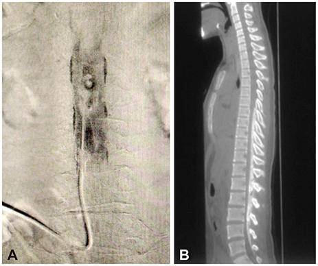
Fig. 1 Spine images in a patient with CSF leakage in and after EBP. A: X-ray photography to confirm the epidural infusion of contrast medium before applying an EBP. B: Sagittal CT image obtained after applying the EBP. Epidural blood mixed with contrast medium is evident from the thoracic sixth to the lumbar fifth vertebral levels. EBP: epidural blood patch.

Fig. 2 Epidural CSF collection (arrows) in MRI fat-suppressed heavily T2-weighted images of patients with spinal CSF leakage. A: Upper cervical vertebral level. B: Lower cervical vertebral level. C: Lower lumber vertebral level.
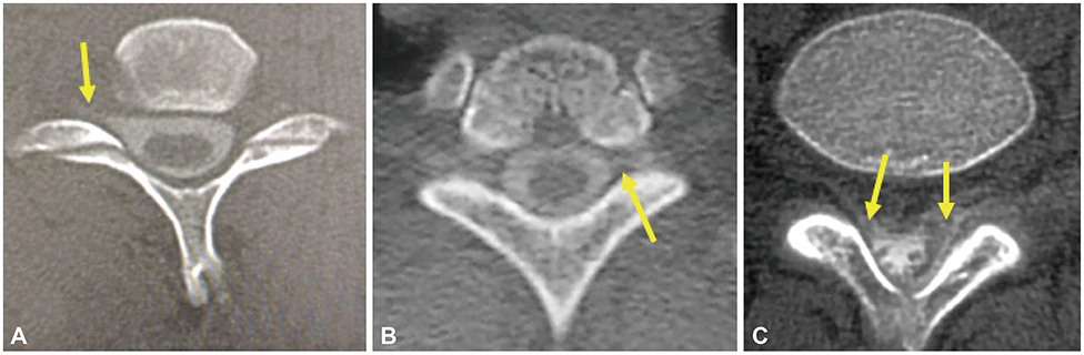
Fig. 3 Epidural CSF leakage (arrows) revealed by CT myelography. A: Epidural CSF leakage at the lower cervical vertebral level. B: Epidural CSF leakage at the upper thoracic vertebral level. C: Epidural CSF leakage at the lumbar vertebral level.
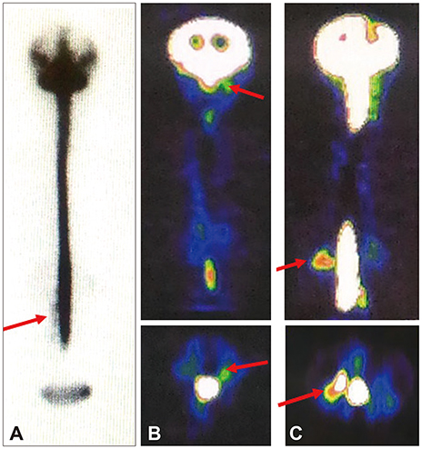
Fig. 4 RI images of patiests with CSF leakage. A: Epidural CSF leakage at the lumbar vertebral level in RI cisternography. B: Epidural CSF leakage at the upper cervical vertebral level in coronal (upper) and axial (lower) SPECT images. C: Epidural CSF leakage at the upper lumbar vertebral level in coronal (upper) and axial (lower) on SPECT images. Leak points of CSF (arrows, A–C). RI: radioisotope.
Reference
-
1. Schievink WI. Spontaneous spinal cerebrospinal fluid leaks and intracranial hypotension. JAMA. 2006; 295:2286–2296.
Article2. Mokri B. Spontaneous low pressure, low CSF volume headaches: spontaneous CSF leaks. Headache. 2013; 53:1034–1053.
Article3. Mokri B, Atkinson JL, Piepgras DG. Absent headache despite CSF volume depletion (intracranial hypotension). Neurology. 2000; 55:1722–1724.
Article4. Schievink WI, Maya MM, Louy C, Moser FG, Tourje J. Diagnostic criteria for spontaneous spinal CSF leaks and intracranial hypotension. AJNR Am J Neuroradiol. 2008; 29:853–856.
Article5. Mokri B, Schievink WI. Headache associated with abnormalities in intracranial structure and function: low cerebrospinal fluid pressure headache. In : Silberstein SD, Lipton RB, Dodick DW, editors. Wolff's Headache and Other Head Pain. New York: Oxford University Press;2000. p. 513–531.6. Horton JC, Fishman RA. Neurovisual findings in the syndrome of spontaneous intracranial hypotension from dural cerebrospinal fluid leak. Ophthalmology. 1994; 101:244–251.
Article7. Brady-McCreery KM, Speidel S, Hussein MA, Coats DK. Spontaneous intracranial hypotension with unique strabismus due to third and fourth cranial neuropathies. Binocul Vis Strabismus Q. 2002; 17:43–48.8. Ferrante E, Savino A, Brioschi A, Marazzi R, Donato MF, Riva M. Transient oculomotor cranial nerves palsy in spontaneous intracranial hypotension. J Neurosurg Sci. 1998; 42:177–179.9. Nowak DA, Rodiek SO, Zinner J, Guhlmann A, Topka H. Broadening the clinical spectrum: unusual presentation of spontaneous cerebrospinal fluid hypovolemia. J Neurosurg. 2003; 98:903–907.
Article10. Hong M, Shah GV, Adams KM, Turner RS, Foster NL. Spontaneous intracranial hypotension causing reversible frontotemporal dementia. Neurology. 2002; 58:1285–1287.
Article11. Wicklund MR, Mokri B, Drubach DA, Boeve BF, Parisi JE, Josephs KA. Frontotemporal brain sagging syndrome: an SIH-like presentation mimicking FTD. Neurology. 2011; 76:1377–1382.
Article12. Schievink WI, Meyer FB, Atkinson JL, Mokri B. Spontaneous spinal cerebrospinal fluid leaks and intracranial hypotension. J Neurosurg. 1996; 84:598–605.
Article13. Pannullo SC, Reich JB, Krol G, Deck MD, Posner JB. MRI changes in intracranial hypotension. Neurology. 1993; 43:919–926.
Article14. Akbar JJ, Luetmer PH, Schwartz KM, Hunt CH, Diehn FE, Eckel LJ. The role of MR myelography with intrathecal gadolinium in localization of spinal CSF leaks in patients with spontaneous intracranial hypotension. AJNR Am J Neuroradiol. 2012; 33:535–540.
Article15. Duffy PJ, Crosby ET. The epidural blood patch. Resolving the controversies. Can J Anaesth. 1999; 46:878–886.
Article16. Sencakova D, Mokri B, McClelland RL. The efficacy of epidural blood patch in spontaneous CSF leaks. Neurology. 2001; 57:1921–1923.
Article17. Karm MH, Choi JH, Kim D, Park JY, Yun HJ, Suh JH. Predictors of the treatment response of spontaneous intracranial hypotension to an epidural blood patch. Medicine (Baltimore). 2016; 95:e3578.
Article18. Wu JW, Hseu SS, Fuh JL, Lirng JF, Wang YF, Chen WT, et al. Factors predicting response to the first epidural blood patch in spontaneous intracranial hypotension. Brain. 2017; 140:344–352.
Article19. Schievink WI. Spontaneous spinal cerebrospinal fluid leaks. Cephalalgia. 2008; 28:1345–1356.
Article20. Mokri B. Headaches caused by decreased intracranial pressure: diagnosis and management. Curr Opin Neurol. 2003; 16:319–326.
Article21. Cho KI, Moon HS, Jeon HJ, Park K, Kong DS. Spontaneous intracranial hypotension: efficacy of radiologic targeting vs blind blood patch. Neurology. 2011; 76:1139–1144.
Article22. Gibson BE, Wedel DJ, Faust RJ, Petersen RC. Continuous epidural saline infusion for the treatment of low CSF pressure headache. Anesthesiology. 1988; 68:789–791.
Article23. Crul BJ, Gerritse BM, Van Dongen RT, Schoonderwaldt HC. Epidural fibrin glue injection stops persistent postdural puncture headache. Anesthesiology. 1999; 91:576–577.
Article24. Franzini A, Messina G, Nazzi V, Mea E, Leone M, Chiapparini L, et al. Spontaneous intracranial hypotension syndrome: a novel speculative physiopathological hypothesis and a novel patch method in a series of 28 consecutive patients. J Neurosurg. 2010; 112:300–306.
Article25. Spelle L, Boulin A, Tainturier C, Visot A, Graveleau P, Pierot L. Neuroimaging features of spontaneous intracranial hypotension. Neuroradiology. 2001; 43:622–627.
Article26. Joo EY, Hwang BY, Kong YG, Lee JH, Hwang BS, Suh JH. Retrospective study of epidural blood patch use for spontaneous intracranial hypotension. Reg Anesth Pain Med. 2015; 40:58–61.
Article27. Moriyama E, Ogawa T, Nishida A, Ishikawa S, Beck H. Quantitative analysis of radioisotope cisternography in the diagnosis of intracranial hypotension. J Neurosurg. 2004; 101:421–426.
Article
- Full Text Links
-
- Actions
-
Cited
- CITED
-
- Close
- Share
-
- Similar articles
-
- Epidural Blood Patches in a Patient With Multi-level Cerebrospinal Fluid Leakage That Was Induced by Spontaneous Intracranial Hypotension
- Spontaneous Intracranial Hypotension Treated with CT-guided Cervical Epidural Blood Patch : A case report
- Detection of Surgery-related Spinal Cerebrospinal Fluid Leakage Using Magnetic Resonance Myelography
- A Clinical Study of Postlumbar-Puncture Headaches
- Spontaneous Intracranial Hypotension Treated with Epidural Blood Patch
