Eccrine Syringofibroadenoma Associated with Bowen's Disease: A Case Report and Review of the Literature
- Affiliations
-
- 1Department of Dermatology, SMG-SNU Boramae Medical Center, Seoul, Korea. sycho@snu.ac.kr
- KMID: 2467202
- DOI: http://doi.org/10.5021/ad.2020.32.1.57
Abstract
- Eccrine syringofibroadenoma (ESFA) is a rare, benign adnexal neoplasm which usually manifests as a solitary nodule on the extremities of elderly patients. Few case reports have described an association between ESFA and carcinomas including squamous cell carcinoma, porocarcinoma, and basal cell carcinoma. A 66-year-old male presented with a pruritic, erythematous brownish solitary plaque with crusted and verrucous surface on the extensor side of the right thigh. The lesion developed 6 to 7 years ago, and had been growing slowly. Biopsy revealed anastomosing epithelial strands which were composed of 2 areas: the upper area consisting of dysplastic cells with prominent nucleoli and abundant mitoses, and the lower area consisting of oval and round cells, and occasionally small luminal ducts. Dysplastic cells comprised almost the entire epidermis but did not invade into the dermis. Benign syringofibroadenomatous lesion surrounded the dysplastic cells in the lowermost portion of the epidermis. Although it is still unclear whether ESFA undergoes malignant transformation or it is a reactive change to carcinoma, complete excision should be performed to prevent malignant transformation with unknown risk.
Keyword
MeSH Terms
Figure
-

Fig. 1 Pruritic solitary mass on the extensor surface of the right thigh. A 3×2 cm sized, well-circumscribed, erythematous brownish plaque with verrucous and partially crusted surface.
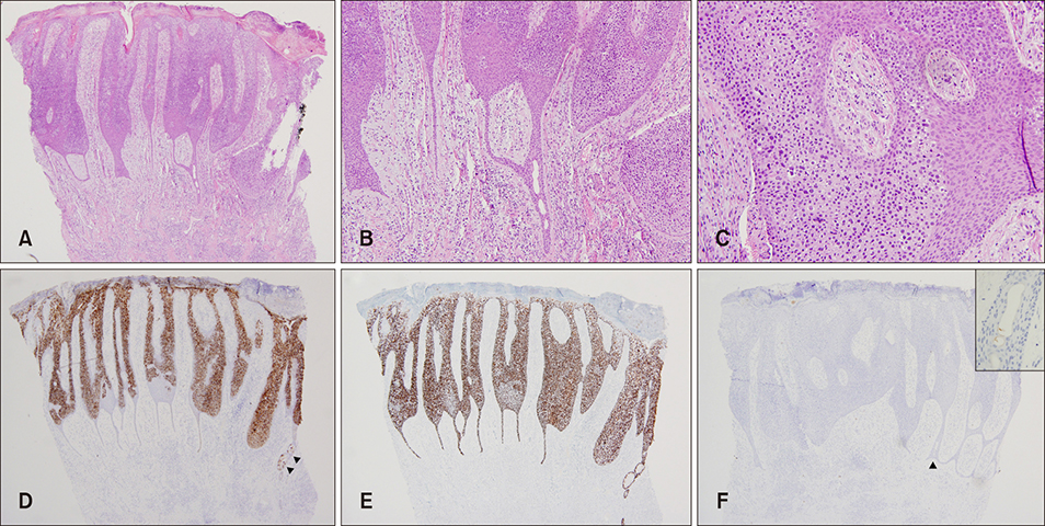
Fig. 2 Histopathology of the solitary mass. (A) Anastomosing epithelial strands extended down to the dermis and were composed of 2 different areas, i.e., dysplastic cells comprising almost the entire epidermis but not invading into the dermis and benign adenomatous lesion immediately surrounding the dysplastic keratinocytes in the lowermost portion of the epidermis (hematoxylin and eosin [H&E], ×40). (B) Epithelial strands were surrounded by mucinous fibrous stroma, and small luminal ducts were embedded within the strands (H&E, ×100). (C) Dysplastic area was composed of atypical cells with prominent nucleoli and abundant mitoses. Adjacent benign adenomatous portion was composed of oval and round cells (H&E, ×200). (D) Epithelial membrane antigen (EMA) was positive in the cytoplasm of dysplastic cells and weakly positive in the ductal structures in the adenomatous lesion (arrowheads) (EMA, ×40). (E) A p63 was positive in the nucleus of dysplastic cells and weakly positive in the adenoma lesion (p63, ×40). (F) Carcinoembryonic antigen (CEA) stain was weakly positive in the ductal structures in the adenoma lesion (arrowhead and inset on the right upper corner) (CEA, ×40).
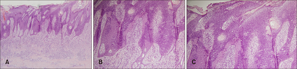
Fig. 3 Histopathology of excised tissues. (A) Epithelial strands formed anastomoses and ductal structures were observed within the epithelial strands extending down into the dermis (hematoxylin and eosin [H&E], ×40). (B, C) Immediately above and adjacent to the syringofibroadenoma lesion, almost the entire epidermis displayed dysplasia with atypical keratinocytes and numerous mitotic figures (H&E, ×100).
Reference
-
1. Bottino CB, Guimarães TF, Gomes FR, D'Acri AM, Lima RB, Martins CJ. Solitary eccrine syringofibroadenoma--case report. An Bras Dermatol. 2015; 90:3 Suppl 1. 235–238.2. Cho E, Lee JD, Cho SH. A case of reactive eccrine syringofibroadenoma. Ann Dermatol. 2011; 23:70–72.
Article3. Fernandez-Flores A, Suarez-Penaranda JM, Halec G, Michael KM, Schmitt M. Study of squamous cell carcinoma associated with syringofibroadenoma for 105 types of human papillomavirus and for all currently known types of polyomaviruses. Appl Immunohistochem Mol Morphol. 2014; 22:e41–e44.
Article4. Duffy KL, Bowen AR, Tristani-Firouzi P, Florell SR, Hadley ML. Eccrine syringofibroadenoma-like change adjacent to a squamous cell carcinoma: potential histologic pitfall in Mohs micrographic surgery. Dermatol Surg. 2009; 35:519–522.
Article5. French LE. Reactive eccrine syringofibroadenoma: an emerging subtype. Dermatology. 1997; 195:309–310.
Article6. Hays JP, Malone CH, Goodwin BP, Wagner RF Jr. Reactive Eccrine syringofibroadenoma associated with basal cell carcinoma: a histologic mimicker of fibroepithelioma of pinkus. Dermatol Surg. 2018; 44:738–740.
Article7. Kacerovska D, Nemcova J, Michal M, Kazakov DV. Eccrine syringofibroadenoma associated with well-differentiated squamous cell carcinoma. Am J Dermatopathol. 2008; 30:572–574.
Article8. González-Serva A, Pró-Rísquez MA, Oliver M, Caruso MG. Syringofibrocarcinoma versus squamous cell carcinoma involving syringofibroadenoma: is there a malignant counterpart of Mascaro's syringofibroadenoma? Am J Dermatopathol. 1997; 19:58–65.
Article9. Schadt CR, Boyd AS. Eccrine syringofibroadenoma with coexistent squamous cell carcinoma. J Cutan Pathol. 2007; 34 Suppl 1:71–74.
Article10. D'Amato MS, Patterson RH, Guccion JG, White JC, Krasnow SH. Porocarcinoma of the heel. A case report with unusual histologic features. Cancer. 1996; 78:751–757.11. Starink TM. Eccrine syringofibroadenoma: multiple lesions representing a new cutaneous marker of the Schöpf syndrome, and solitary nonhereditary tumors. J Am Acad Dermatol. 1997; 36:569–576.
Article12. Lele SM, Gloster ES, Heilman ER, Chen PC, Chen CK, Anzil AP, et al. Eccrine syringofibroadenoma surrounding a squamous cell carcinoma: a case report. J Cutan Pathol. 1997; 24:193–196.
Article13. Katane M, Akiyama M, Ohnishi T, Watanabe S, Matsuo I. Carcinomatous transformation of eccrine syringofibroadenoma. J Cutan Pathol. 2003; 30:211–214.
Article14. Bjarke T, Ternesten-Bratel A, Hedblad M, Rausing A. Carcinoma and eccrine syringofibroadenoma: a report of five cases. J Cutan Pathol. 2003; 30:382–392.
Article15. Cho HS, Kim WH, Kim CW, Kim KH, Kim KJ. A case of eccrine syringofibroadenoma associated with squamous cell carcinoma. Korean J Dermatol. 2005; 43:1635–1638.16. Watanabe R, Mikami M, Kitami A, Akiyama M, Hirohiko S, Iijima M, et al. Eccrine porocarcinoma accompanied by eccrine syringofibroadenoma-like histology. Skin Cancer. 2007; 22:62–67.
Article17. Crowson AN, Magro CM, Mihm MC. Malignant adnexal neoplasms. Mod Pathol. 2006; 19 Suppl 2:S93–S126.
Article18. Calonje E, Lazar A, Mckee PH. McKee's pathology of the skin: with clinical correlations. 4th ed. Edinburgh: Elsevier/Saunders;2012.
