Detection of peri-implant bone defects using cone-beam computed tomography and digital periapical radiography with parallel and oblique projection
- Affiliations
-
- 1Dental Sciences Research Center, Department of Periodontics, School of Dentistry, Guilan University of Medical Sciences, Rasht, Iran.
- 2Dental Sciences Research Center, Department of Maxillofacial Radiology, School of Dentistry, Guilan University of Medical Sciences, Rasht, Iran. ngrkhosravi@yahoo.com
- 3Department of Maxillofacial Radiology, School of Dentistry, Guilan University of Medical Sciences, Rasht, Iran.
- KMID: 2466548
- DOI: http://doi.org/10.5624/isd.2019.49.4.265
Abstract
- PURPOSE
To compare the diagnostic accuracy of cone-beam computed tomography (CBCT) with that of parallel (PPA) and oblique projected periapical (OPA) radiography for the detection of different types of peri-implant bone defects.
MATERIALS AND METHODS
Forty implants inserted into bovine rib blocks were used. Thirty had standardized bone defects (10 each of angular, fenestration, and dehiscence defects), and 10 were defect-free controls. CBCT, PPA, and OPA images of the samples were acquired. The images were evaluated twice by each of 2 blinded observers regarding the presence or absence and the type of the defects. The area under the receiver operating characteristic curve (AUC), sensitivity, and specificity were determined for each radiographic technique. The 3 modalities were compared using the Fisher exact and chi-square tests, with P<0.05 considered as statistical significance.
RESULTS
High inter-examiner reliability was observed for the 3 techniques. Angular defects were detected with high sensitivity and specificity by all 3 modalities. CBCT and OPA showed similar AUC and sensitivity in the detection of fenestration defects. In the identification of dehiscence defects, CBCT showed the highest sensitivity, followed by OPA and PPA, respectively. CBCT and OPA had a significantly greater ability than PPA to detect fenestration and dehiscence defects (P<0.05).
CONCLUSION
The application of OPA radiography in addition to routine PPA imaging as a radiographic follow-up method for dental implantation greatly enhances the visualization of fenestration and dehiscence defects. CBCT properly depicted all defect types studied, but it involves a relatively high dose of radiation and cost.
MeSH Terms
Figure
-

Fig. 1 Peri-implant bone defects created in bovine bone blocks. A. Fenestration defect. B. Dehiscence defect. C. Three-wall defect. D. Two-wall defect.
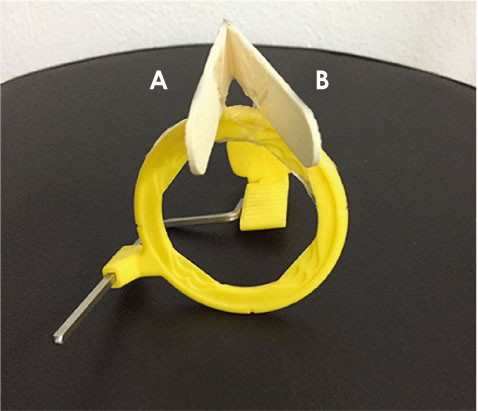
Fig. 2 Tongue depressors attached to the holder device for the acquisition of parallel (A) and oblique (B) periapical radiographs.
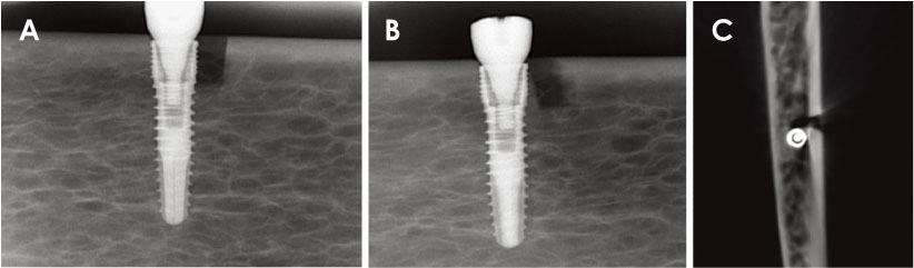
Fig. 3 Radiographic images of a 2-wall angular defect. A. Parallel periapical radiograph. B. Oblique periapical radiograph. C. Axial cone-beam computed tomographic image.
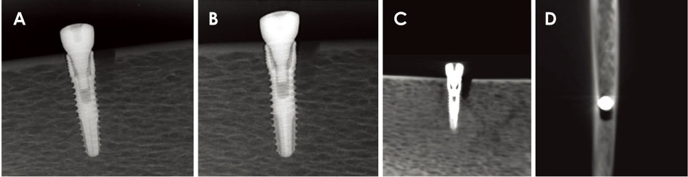
Fig. 4 Radiographic images of a 3-wall angular defect. A. Parallel periapical radiograph. B. Oblique periapical radiograph. C. Tangential cone-beam computed tomographic image. D. Axial cone-beam computed tomographic image.
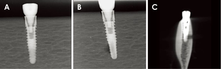
Fig. 5 Radiographic images of a fenestration defect. A. Parallel periapical radiograph. B. Oblique periapical radiograph. C. Cross-sectional cone-beam computed tomographic image.
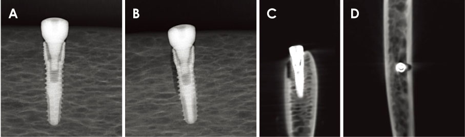
Fig. 6 Radiographic images of a dehiscence defect. A. Parallel periapical radiograph. B. Oblique periapical radiograph. C. Cross-sectional cone-beam computed tomographic image. D. Axial cone-beam computed tomographic image.
Reference
-
1. Ding Q, Zhang L, Geraets W, Wu W, Zhou Y, Wismeijer D, et al. Association between peri-implant bone morphology and marginal bone loss: a retrospective study on implant-supported mandibular overdentures. Int J Oral Maxillofac Implants. 2017; 32:147–155.
Article2. Kamburoğlu K, Murat S, Kılıç C, Yüksel S, Avsever H, Farman A, et al. Accuracy of CBCT images in the assessment of buccal marginal alveolar peri-implant defects: effect of field of view. Dentomaxillofac Radiol. 2014; 43:20130332.
Article3. Dave M, Davies J, Wilson R, Palmer R. A comparison of cone beam computed tomography and conventional periapical radiography at detecting peri-implant bone defects. Clin Oral Implants Res. 2012; 24:671–678.
Article4. Bagis N, Kolsuz ME, Kursun S, Orhan K. Comparison of intraoral radiography and cone-beam computed tomography for the detection of periodontal defects: an in vitro study. BMC Oral Health. 2015; 15:64.
Article5. Bohner LO, Mukai E, Oderich E, Porporatti AL, Pacheco-Pereira C, Tortamano P, et al. Comparative analysis of imaging techniques for diagnostic accuracy of peri-implant bone defects: a meta-analysis. Oral Surg Oral Med Oral Pathol Oral Radiol. 2017; 124:432–440.
Article6. Hilgenfeld T, Juerchott A, Deisenhofer UK, Krisam J, Rammelsberg P, Heiland S, et al. Accuracy of cone-beam computed tomography, dental magnetic resonance imaging, and intraoral radiography for detecting peri-implant bone defects at single zirconia implants - an in vitro study. Clin Oral Implants Res. 2018; 29:922–930.7. Saberi BV, Khosravifard N, Mohtavipour T, Khaksari F, Abbasi S, Shahmalakpoor A. Entrance skin dose of the thyroid gland area following exposure with different protocols of two panoramic and cone-beam computed tomography devices. J Oral Maxillofac Radiol. 2019; 7:6–11.
Article8. Khojastepour L, Haghnegahdar A, Khosravifard N. Role of sinonasal anatomic variations in the development of maxillary sinusitis: a cone beam CT analysis. Open Dent J. 2017; 11:367–374.
Article9. de-Azevedo-Vaz SL, Peyneau PD, Ramirez-Sotelo LR, Vasconcelos Kde F, Campos PS, Haiter-Neto F. Efficacy of a cone beam computed tomography metal artifact reduction algorithm for the detection of peri-implant fenestrations and dehiscences. Oral Surg Oral Med Oral Pathol Oral Radiol. 2016; 121:550–556.
Article10. de-Azevedo-Vaz SL, Vasconcelos Kde F, Neves FS, Melo SL, Campos PS, Haiter-Neto F. Detection of periimplant fenestration and dehiscence with the use of two scan modes and the smallest voxel sizes of a cone-beam computed tomography device. Oral Surg Oral Med Oral Pathol Oral Radiol. 2013; 115:121–127.
Article11. Liedke GS, Spin-Neto R, da Silveira HE, Schropp L, Stavropoulos A, Wenzel A. Factors affecting the possibility to detect buccal bone condition around dental implants using cone beam computed tomography. Clin Oral Implants Res. 2017; 28:1082–1088.
Article12. Mikolajczak T, Wilk G. The diagnostic value of oblique technique for periapical radiography and its usefulness in endodontic treatment. Ann Acad Med Stetin. 2008; 54:94–98.13. Pinheiro LR, Scarfe WC, Augusto de Oliveira Sales M, Gaia BF, Cortes AR, Cavalcanti MG. Effect of cone-beam computed tomography field of view and acquisition frame on the detection of chemically simulated peri-implant bone loss in vitro. J Periodontol. 2015; 86:1159–1165.
Article14. Salvi GE, Cosgarea R, Sculean A. Prevalence and mechanisms of peri-implant diseases. J Dent Res. 2017; 96:31–37.
Article15. Khoshkam V, Chan HL, Lin GH, MacEachern MP, Monje A, Suarez F, et al. Reconstructive procedures for treating peri-implantitis: a systematic review. J Dent Res. 2013; 92(12 Suppl):131s–138s.16. Silveira-Neto N, Flores ME, De Carli JP, Costa MD, Matos FS, Paranhos LR, et al. Peri-implant assessment via cone beam computed tomography and digital periapical radiography: an ex vivo study. Clinics (Sao Paulo). 2017; 72:708–713.
Article17. Ritter L, Elger MC, Rothamel D, Fienitz T, Zinser M, Schwarz F, et al. Accuracy of peri-implant bone evaluation using cone beam CT, digital intra-oral radiographs and histology. Dentomaxillofac Radiol. 2014; 43:20130088.
Article18. Schwarz F, Sahm N, Schwarz K, Becker J. Impact of defect configuration on the clinical outcome following surgical regenerative therapy of peri-impantitis. J Clin Periodontol. 2010; 37:449–455.19. Mengel R, Kruse B, Flores-de-Jacoby L. Digital volume tomography in the diagnosis of peri-implant defects: an in vitro study on native pig mandibles. J Periodontol. 2006; 77:1234–1241.
Article20. Sirin Y, Horasan S, Yaman D, Basegmez C, Tanyel C, Aral A, et al. Detection of crestal radiolucencies around dental implants: an in vitro experimental study. J Oral Maxillofac Surg. 2012; 70:1540–1550.
Article21. Kühl S, Zürcher S, Zitzmann NU, Filippi A, Payer M, Dagassan-Berndt D. Detection of peri-implant bone defects with different radiographic techniques - a human cadaver study. Clin Oral Implants Res. 2016; 27:529–534.
Article22. Eskandarloo A, Saati S, Ardakani MP, Jamalpour M, Gholi Mezerji NM, Akheshteh V. Diagnostic accuracy of three cone beam computed tomography systems and periapical radiography for detection of fenestration around dental implants. Contemp Clin Dent. 2018; 9:367–381.
- Full Text Links
-
- Actions
-
Cited
- CITED
-
- Close
- Share
-
- Similar articles
-
- Evaluation of peri-implant bone defects on cone-beam computed tomography and the diagnostic accuracy of detecting these defects on panoramic images
- Evaluation of bone quality in alveolar crest obscured by dental implants: A pilot study by densitometric digital analysis in mandibular bone specimen
- Detection of maxillary second molar with two palatal roots using cone beam computed tomography: a case report
- Commentary on "Reliability of two different presurgical preparation methods for implant dentistry based on panoramic radiography and cone-beam computed tomography in cadavers"
- A comparative study of cone-beam computed tomography and digital periapical radiography in detecting mandibular molars root perforations
