Imaging Sci Dent.
2014 Jun;44(2):115-119. 10.5624/isd.2014.44.2.115.
A comparative study of cone-beam computed tomography and digital periapical radiography in detecting mandibular molars root perforations
- Affiliations
-
- 1Department of Oral and Maxillofacial Radiology, Dental Material Research Center, Dental Faculty, Babol University of Medical Sciences, Babol, Iran. dr.abbaszadeh1@gmail.com
- 2Department of Endodontics, Dental Material Research Center, Dental Faculty, Babol University of Medical Sciences, Babol, Iran.
- 3Non-Communicable Pediatric Diseases Research Center, Babol University of Medical Sciences, Babol, Iran.
- KMID: 1799592
- DOI: http://doi.org/10.5624/isd.2014.44.2.115
Abstract
- PURPOSE
The aim of this in vitro study was to determine the sensitivity and specificity of cone-beam computed tomography (CBCT) and digital periapical radiography in the detection of mesial root perforations of mandibular molars.
MATERIALS AND METHODS
In this in vitro study, 48 mandibular molars were divided into 4 groups. First, the mesial canals of all the 48 teeth were endodontically prepared. In 2 groups (24 teeth each), the roots were axially perforated in the mesiolingual canal 1-3 mm below the furcation region, penetrating the root surface ("root perforation"). Then, in one of these 2 groups, the mesial canals were filled with gutta-percha and AH26 sealer. Mesial canals in one of the other 2 groups without perforation (control groups) were filled with the same materials. The CBCT and periapical radiographs with 3 different angulations were evaluated by 2 oral and maxillofacial radiologists. The specificity and sensitivity of the two methods were calculated, and P<0.05 was considered significant.
RESULTS
The sensitivity and specificity of CBCT scans in the detection of obturated root canal perforations were 79% and 96%, respectively, and in the case of three-angled periapical radiographs, they were 92% and 100%, respectively. In non-obturated root canals, the sensitivity and specificity of CBCT scans in perforation detection were 92% and 100%, respectively, and for three-angled periapical radiographs, they were 50% and 96%, respectively.
CONCLUSION
For perforation detection in filled-root canals, periapical radiography with three different horizontal angulations would be trustworthy, but it is recommended that CBCT be used for perforation detection before obturating root canals.
MeSH Terms
Figure
-
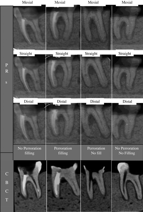
Fig. 1 Periapical radiographs and cone-beam CT scans show the groups of perforated root canals with obturation and no obturation, and the control group.
Cited by 1 articles
-
The effect of metal artifacts on the identification of vertical root fractures using different fields of view in cone-beam computed tomography
Ehsan Moudi, Sina Haghanifar, Zahrasadat Madani, Ali Bijani, Zeynab Sadat Nabavi
Imaging Sci Dent. 2015;45(3):147-151. doi: 10.5624/isd.2015.45.3.147.
Reference
-
1. Ng YL, Mann V, Gulabivala K. A prospective study of the factors affecting outcomes of nonsurgical root canal treatment: part 1: periapical health. Int Endod J. 2011; 44:583–609.
Article2. Tsesis I, Fuss Z. Diagnosis and treatment of accidental root perforations. Endod Topics. 2006; 13:95–107.
Article3. Tsesis I, Rosenberg E, Faivishevsky V, Kfir A, Katz M, Rosen E. Prevalence and associated periodontal status of teeth with root perforation: a retrospective study of 2,002 patients' medical records. J Endod. 2010; 36:797–800.
Article4. Alhadainy HA. Root perforations. A review of literature. Oral Surg Oral Med Oral Pathol. 1994; 78:368–374.5. Shemesh H, Cristescu RC, Wesselink PR, Wu MK. The use of cone-beam computed tomography and digital periapical radiographs to diagnose root perforations. J Endod. 2011; 37:513–516.
Article6. Kamburoglu K, Kursun S, Yuksel S, Oztas B. Observer ability to detect ex vivo simulated internal or external cervical root resorption. J Endod. 2011; 37:168–175.7. Hassan B, Metska ME, Ozok AR, van der Stelt P, Wesselink PR. Detection of vertical root fractures in endodontically treated teeth by a cone beam computed tomography scan. J Endod. 2009; 35:719–722.
Article8. Scarfe WC, Levin MD, Gane D, Farman AG. Use of cone beam computed tomography in endodontics. Int J Dent. 2009; 2009:634567.
Article9. Kajan ZD, Taromsari M. Value of cone beam CT in detection of dental root fractures. Dentomaxillofac Radiol. 2012; 41:3–10.
Article10. Patel S, Dawood A, Wilson R, Horner K, Mannocci F. The detection and management of root resorption lesions using intraoral radiography and cone beam computed tomography - an in vivo investigation. Int Endod J. 2009; 42:831–838.11. Kamburoğlu K, Murat S, Yüksel SP, Cebeci AR, Horasan S. Detection of vertical root fracture using cone-beam computerized tomography: an in vitro assessment. Oral Surg Oral Med Oral Pathol Oral Radiol Endod. 2010; 109:e74–e81.
Article12. Schneider SW. A comparison of canal preparations in straight and curved root canals. Oral Surg Oral Med Oral Pathol. 1971; 32:271–275.
Article13. Bechara B, Alex McMahan C, Moore WS, Noujeim M, Teixeira FB, Geha H. Cone beam CT scans with and without artefact reduction in root fracture detection of endodontically treated teeth. Dentomaxillofac Radiol. 2013; 42:20120245.
Article14. Bueno MR, Estrela C, De Figueiredo JA, Azevedo BC. Mapreading strategy to diagnose root perforations near metallic intracanal posts by using cone-beam computed tomography. J Endod. 2011; 37:85–90.15. Regan JD, Witherspoon DE, Foyle D. Surgical repair of root and tooth perforations. Endod Topics. 2005; 11:152–178.
Article16. Patel S, Dawood A, Ford TP, Whaites E. The potential applications of cone beam computed tomography in the management of endodontic problems. Int Endod J. 2007; 40:818–830.
Article17. Bernardes RA, de Paulo RS, Pereira LO, Duarte MA, Ordinola-Zapata R, de Azevedo JR. Comparative study of cone beam computed tomography and intraoral periapical radiographs in diagnosis of lingual-simulated external root resorptions. Dent Traumatol. 2012; 28:268–272.
Article
- Full Text Links
-
- Actions
-
Cited
- CITED
-
- Close
- Share
-
- Similar articles
-
- Reliability of panoramic radiography in predicting proximity of third molars to the mandibular canal: A comparison using cone-beam computed tomography
- Assessment of the relationship between the mandibular third molar and the mandibular canal using panoramic radiograph and cone beam computed tomography
- Detection of maxillary second molar with two palatal roots using cone beam computed tomography: a case report
- Observation of mandibular second molar roots and root canal morphology using dental cone-beam computed tomography
- Comparison of cone beam computed tomography and conventional panoramic radiography in assessing the topographic relationship between the mandibular canal and impacted third molars
