Progressive Optic Disc Tilt in Young Myopic Glaucomatous Eyes
- Affiliations
-
- 1Department of Ophthalmology, Asan Medical Center, University of Ulsan College of Medicine, Seoul, Korea. sungeye@gmail.com
- 2Division of Biostatistics, Center for Medical Research and Information, Asan Medical Center, University of Ulsan College of Medicine, Seoul, Korea.
- KMID: 2465132
- DOI: http://doi.org/10.3341/kjo.2019.0069
Abstract
- PURPOSE
To explore the progressive change and associated factors of optic disc tilt in young myopic glaucomatous eyes by analyzing long-term follow-up data.
METHODS
Optic disc images were obtained from spectral-domain optical coherence tomography enhanced depth imaging from at least five different visits. At each visit, the disc tilt angle (DTA), defined as the angle between the Bruch's membrane opening plane and the optic canal plane, was estimated at the central frame that passes through the optic disc. Glaucoma progression was assessed on the basis of changes noted on serial optic disc and retinal nerve fiber layer photographs or changes in the visual field (VF). A linear mixed effect model was used to assess the influence of parameters (age, sex, baseline and follow-up intraocular pressure, retinal nerve fiber layer thickness, VF mean deviation, axial length, central corneal thickness), and presence of glaucomatous progression upon DTA change.
RESULTS
A total of 26 eyes of 26 young myopic primary open-angle glaucoma patients (axial length >24.0 mm; mean age, 25.1 ± 4.0 years; mean follow-up, 3.3 ± 0.9 years) were included. DTA was 7.0 ± 3.4 degrees at baseline and 8.3 ± 3.8 degrees at last visit, which represents a significant difference (p < 0.001). Worse VF mean deviation (p < 0.001) and longer axial length (p = 0.006) were significantly associated with DTA increase.
CONCLUSIONS
Young myopic glaucomatous eyes showed progressive optic disc tilting. Progressive optic disc tilting in young myopic glaucomatous eyes may be related to either continuous axial myopic shift or glaucomatous structural change.
Keyword
MeSH Terms
Figure
-
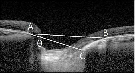
Fig. 1 Measurement of lamina cribrosa depth and disc tilt angle. Bruch's membrane opening (BMO, points A and B) was defined by the proximal tips of the Bruch's membrane. A line connecting the BMO on each side (line AB), was regarded as the BMO plane. The disc tilt angle was defined as the angle between the BMO plane and the optic canal plane (line AC) that passes through the optic disc (θ).

Fig. 2 Disc tilt angle change over time in all eyes. Redline indicates eyes which show glaucomatous progression while blue line indicates stable eyes.

Fig. 3 Representative case example is shown. Upper images were obtained at baseline examination, and lower images were obtained 39 months later from baseline. (A,B) A 28-year-old woman had progressive tilting in her optic disc (baseline, 9.3 degrees; 39 months later, 10.4 degrees) and deepening of the lamina cribrosa (baseline, 359.9 microns; 39 months later, 366.9 microns). (C) Her visual field and retinal nerve fiber layer photographs showed rapid progression.

Fig. 4 Representative case example is shown. Upper images were obtained at baseline examination and lower images were obtained 52 months later from baseline. (A,B) A 30-year-old man showed progressive tilting in his optic disc (baseline, 8.6 degrees; 52 months later, 11.7 degrees and deepening of lamina cribrosa baseline, 351.5 micron; 52 months later, 380.7 micron), but (C) there was no evidence of glaucomatous progression either in the structural or functional test.
Reference
-
1. Marcus MW, de Vries MM, Junoy Montolio FG, Jansonius NM. Myopia as a risk factor for open-angle glaucoma: a systematic review and meta-analysis. Ophthalmology. 2011; 118:1989–1994.2. Wong TY, Foster PJ, Hee J, et al. Prevalence and risk factors for refractive errors in adult Chinese in Singapore. Invest Ophthalmol Vis Sci. 2000; 41:2486–2494.3. Chon B, Qiu M, Lin SC. Myopia and glaucoma in the South Korean population. Invest Ophthalmol Vis Sci. 2013; 54:6570–6577.4. Lee JE, Sung KR, Park JM, et al. Optic disc and peripapillary retinal nerve fiber layer characteristics associated with glaucomatous optic disc in young myopia. Graefes Arch Clin Exp Ophthalmol. 2017; 255:591–598.5. Yun SC, Hahn IK, Sung KR, et al. Lamina cribrosa depth according to the level of axial length in normal and glaucomatous eyes. Graefes Arch Clin Exp Ophthalmol. 2015; 253:2247–2253.6. Wu Z, Lin C, Crowther M, et al. Impact of rates of change of lamina cribrosa and optic nerve head surface depths on visual field progression in glaucoma. Invest Ophthalmol Vis Sci. 2017; 58:1825–1833.7. Chung HS, Sung KR, Lee JY, Na JH. Lamina cribrosa-related parameters assessed by optical coherence tomography for prediction of future glaucoma progression. Curr Eye Res. 2016; 41:806–813.8. Lee EJ, Kim TW, Kim M, Kim H. Influence of lamina cribrosa thickness and depth on the rate of progressive retinal nerve fiber layer thinning. Ophthalmology. 2015; 122:721–729.9. Radius RL, Anderson DR. Rapid axonal transport in primate optic nerve: distribution of pressure-induced interruption. Arch Ophthalmol. 1981; 99:650–654.10. Quigley HA, Anderson DR. Distribution of axonal transport blockade by acute intraocular pressure elevation in the primate optic nerve head. Invest Ophthalmol Vis Sci. 1977; 16:640–644.11. Quigley HA, Addicks EM, Green WR, Maumenee AE. Optic nerve damage in human glaucoma. II. The site of injury and susceptibility to damage. Arch Ophthalmol. 1981; 99:635–649.12. Heijl A, Leske MC, Bengtsson B, et al. Measuring visual field progression in the Early Manifest Glaucoma Trial. Acta Ophthalmol Scand. 2003; 81:286–293.13. Kim CS, Seong GJ, Lee NH, et al. Prevalence of primary open-angle glaucoma in central South Korea the Namil study. Ophthalmology. 2011; 118:1024–1030.14. Kim YJ, Yun SC, Na JH, et al. Glaucoma progression in eyes with a history of refractive corneal surgery. Invest Ophthalmol Vis Sci. 2012; 53:4485–4489.15. Kim TW, Kim M, Weinreb RN, et al. Optic disc change with incipient myopia of childhood. Ophthalmology. 2012; 119:21–26.16. Kim M, Choung HK, Lee KM, et al. Longitudinal changes of optic nerve head and peripapillary structure during childhood myopia progression on OCT: Boramae myopia cohort study report 1. Ophthalmology. 2018; 125:1215–1223.17. Song MK, Sung KR, Shin JW, et al. Progressive change in peripapillary atrophy in myopic glaucomatous eyes. Br J Ophthalmol. 2018; 102:1527–1532.18. Han S, Sung KR, Park J, et al. Sub-classification of myopic glaucomatous eyes according to optic disc and peripapillary features. PLoS One. 2017; 12:e0181841.19. Lee JE, Sung KR, Lee JY, Park JM. Implications of optic disc tilt in the progression of primary open-angle glaucoma. Invest Ophthalmol Vis Sci. 2015; 56:6925–6931.20. Doshi A, Kreidl KO, Lombardi L, et al. Nonprogressive glaucomatous cupping and visual field abnormalities in young Chinese males. Ophthalmology. 2007; 114:472–479.21. Kwon J, Sung KR, Park JM. Myopic glaucomatous eyes with or without optic disc shape alteration: a longitudinal study. Br J Ophthalmol. 2017; 101:1618–1622.
- Full Text Links
-
- Actions
-
Cited
- CITED
-
- Close
- Share
-
- Similar articles
-
- Comparison of Optic Nerve Head Measurements by Optical Coherence Tomography and Confocal Scanning Laser Ophthalmoscopy
- Comparison between Optic Disc Torsion of Fellow Eyes in Unilateral Normal-tension Glaucoma and Normal Eyes
- Development of Optic Disc Torsion in Children
- Relationship between Optic Disc Parameters and Myopic Refractive Errors in Myopia
- Optic disc of the myopic eye: relationship between refractive errors and morphometric characteristics
