Effect of archwire stiffness and friction on maxillary posterior segment displacement during anterior segment retraction: A three-dimensional finite element analysis
- Affiliations
-
- 1Department of Orthodontics, College of Dentistry, Yonsei University, Seoul, Korea. orthojn@yuhs.ac
- 2Division of Orthodontics, Department of Dentistry, Yeouido St. Mary's Hospital, College of Medicine, The Catholic University of Korea, Seoul, Korea.
- 3Institute of Craniofacial Deformity, College of Dentistry, Yonsei University, Seoul, Korea.
- KMID: 2464202
- DOI: http://doi.org/10.4041/kjod.2019.49.6.393
Abstract
OBJECTIVE
Sliding mechanics using orthodontic miniscrews is widely used to stabilize the anchorage during extraction space closure. However, previous studies have reported that both posterior segment displacement and anterior segment displacement are possible, depending on the mechanical properties of the archwire. The present study aimed to investigate the effect of archwire stiffness and friction change on the displacement pattern of the maxillary posterior segment during anterior segment retraction with orthodontic miniscrews in sliding mechanics.
METHODS
A three-dimensional finite element model was constructed. The retraction point was set at the archwire level between the lateral incisor and canine, and the orthodontic miniscrew was located at a height of 8 mm from the archwire between the second premolar and first molar. Archwire stiffness was simulated with rectangular stainless steel wires and a rigid body was used as a control. Various friction levels were set for the surface contact model. Displacement patterns for the posterior and anterior segments were compared between the conditions.
RESULTS
Both the anterior and posterior segments exhibited backward rotation, regardless of archwire stiffness or friction. Among the conditions tested in this study, the least undesirable rotation was found with low archwire stiffness and low friction.
CONCLUSIONS
Posterior segment displacement may be unavoidable but reducing the stiffness and friction of the main archwire may minimize unwanted rotations during extraction space closure.
MeSH Terms
Figure
-
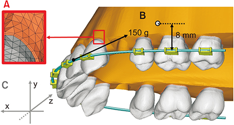
Figure 1 Three-dimensional finite element model and experimental conditions. A, Finite element method model of periodontal ligament. B, Position of orthodontic miniscrews and direction of the retraction force. C, Coordinate system. x-axis: (+) anterior, (−) posterior direction; y-axis: (+) superior, (−) inferior direction; z-axis: (+) buccal, (−) palatal direction.

Figure 2 Landmarks used for the assessment of displacement. Red dots indicate reference points for the crown and root of each tooth. CI, Central incisor; PM2, second premolar; M1, first molar; M2, second molar.
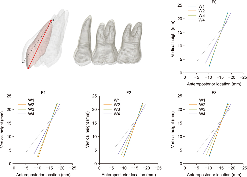
Figure 3 Displacement patterns of the central incisor according to archwire stiffness at each given level of friction. Dotted line, initial position. Solid line, position after movement (50× magnification). Positive value, anterior direction of the x-axis and apical direction of the y-axis. W1, 0.016 × 0.022-inch (in) flexible stainless steel (SS); W2, 0.017 × 0.025-in flexible SS; W3, 0.019 × 0.025-in flexible SS; W4, rigid body. F0, µ = 0.0; F1, µ = 0.1; F2, µ = 0.2; F3, µ = 0.3.

Figure 4 Displacement patterns of posterior teeth according to archwire stiffness at each given level of friction. A, Tooth axis displacement patterns (1,000× magnification). B, Posterior occlusal plane displacement patterns (1,500× magnification). Dotted (gray) line, initial position. Solid line, position after displacement. Positive value, anterior direction of the x-axis and apical direction of the y-axis. See Figures 2 and 3 for definitions of each landmark or measurement.
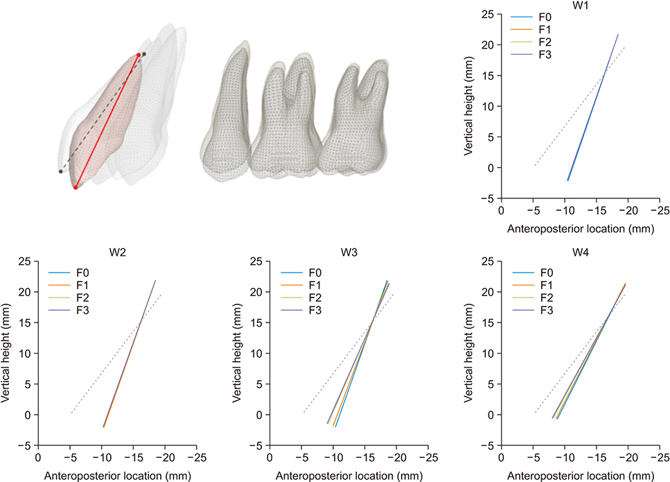
Figure 5 Displacement patterns of the central incisor according to friction levels at each given level of archwire stiffness. Dotted line, initial position. Solid line, position after movement (50× magnification). Positive value, anterior direction of the x-axis and apical direction of the y-axis. See Figure 3 for definitions of each landmark or measurement.
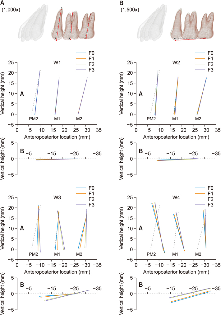
Figure 6 Displacement patterns of posterior teeth according to friction levels at each given level of archwire stiffness. A, Tooth axis displacement patterns (1,000× magnification). B, Posterior occlusal plane displacement patterns (1,500× magnification). Dotted (gray) line, initial position. Solid line, position after displacement. Positive value, anterior direction of the x-axis and apical direction of the y-axis. See Figures 2 and 3 for definitions of each landmark or measurement.
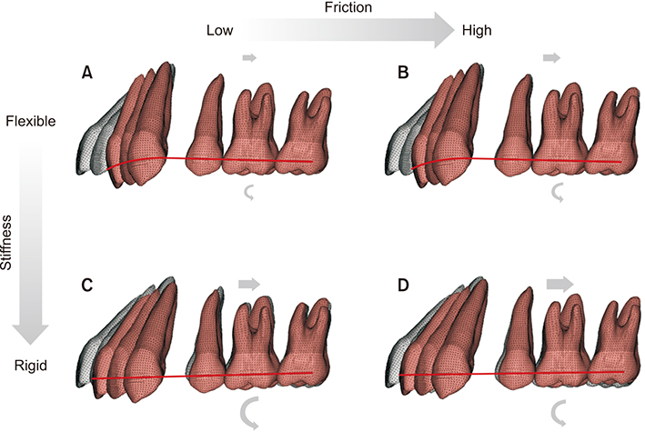
Figure 7 Displacement patterns resulting from varying levels of archwire stiffness and friction. Relatively low archwire stiffness led to less displacement of the posterior segment compared to that observed with high archwire stiffness across friction levels. A, Flexible archwire with low friction. B, Flexible archwire with high friction. C, Rigid archwire with low friction. D, Rigid archwire with high friction (gray color, original tooth position; brown color, tooth position after displacement).
Reference
-
1. Upadhyay M, Yadav S, Nagaraj K, Patil S. Treatment effects of mini-implants for en-masse retraction of anterior teeth in bialveolar dental protrusion patients: a randomized controlled trial. Am J Orthod Dentofacial Orthop. 2008; 134:18–29.e1.
Article2. Song JW, Lim JK, Lee KJ, Sung SJ, Chun YS, Mo SS. Finite element analysis of maxillary incisor displacement during en-masse retraction according to orthodontic mini-implant position. Korean J Orthod. 2016; 46:242–252.
Article3. Bechtold TE, Kim JW, Choi TH, Park YC, Lee KJ. Distalization pattern of the maxillary arch depending on the number of orthodontic miniscrews. Angle Orthod. 2013; 83:266–273.
Article4. Upadhyay M, Yadav S, Patil S. Mini-implant anchorage for en-masse retraction of maxillary anterior teeth: a clinical cephalometric study. Am J Orthod Dentofacial Orthop. 2008; 134:803–810.
Article5. Kojima Y, Kawamura J, Fukui H. Finite element analysis of the effect of force directions on tooth movement in extraction space closure with miniscrew sliding mechanics. Am J Orthod Dentofacial Orthop. 2012; 142:501–508.
Article6. Kojima Y, Fukui H. Numeric simulations of en-masse space closure with sliding mechanics. Am J Orthod Dentofacial Orthop. 2010; 138:702.e1–702.e6. discussion 702–4.
Article7. Tominaga JY, Ozaki H, Chiang PC, Sumi M, Tanaka M, Koga Y, et al. Effect of bracket slot and archwire dimensions on anterior tooth movement during space closure in sliding mechanics: a 3-dimensional finite element study. Am J Orthod Dentofacial Orthop. 2014; 146:166–174.
Article8. Moore JC, Waters NE. Factors affecting tooth movement in sliding mechanics. Eur J Orthod. 1993; 15:235–241.
Article9. Sung SJ, Baik HS, Moon YS, Yu HS, Cho YS. A comparative evaluation of different compensating curves in the lingual and labial techniques using 3D FEM. Am J Orthod Dentofacial Orthop. 2003; 123:441–450.
Article10. Lee KJ, Park YC, Hwang CJ, Kim YJ, Choi TH, Yoo HM, et al. Displacement pattern of the maxillary arch depending on miniscrew position in sliding mechanics. Am J Orthod Dentofacial Orthop. 2011; 140:224–232.
Article11. Jung MH, Kim TW. Biomechanical considerations in treatment with miniscrew anchorage. Part 1: the sagittal plane. J Clin Orthod. 2008; 42:79–83.12. Kojima Y, Fukui H. A finite element simulation of initial movement, orthodontic movement, and the centre of resistance of the maxillary teeth connected with an archwire. Eur J Orthod. 2014; 36:255–261.
Article13. Wakabayashi N, Ona M, Suzuki T, Igarashi Y. Nonlinear finite element analyses: advances and challenges in dental applications. J Dent. 2008; 36:463–471.
Article14. Chong DR, Jang YJ, Chun YS, Jung SH, Lee SK. The evaluation of rotational movements of maxillary posterior teeth using three dimensional images in cases of extraction of maxillary first premolar. Korean J Orthod. 2005; 35:451–458.15. Andrews LF. The six keys to normal occlusion. Am J Orthod. 1972; 62:296–309.
Article16. Coolidge ED. The thickness of the human periodontal membrane. J Am Dent Assoc. 1937; 24:1260–1270.
Article17. Andrews LF. The straight-wire appliance. Explained and compared. J Clin Orthod. 1976; 10:174–195.18. Papageorgiou SN, Keilig L, Hasan I, Jäger A, Bourauel C. Effect of material variation on the biomechanical behaviour of orthodontic fixed appliances: a finite element analysis. Eur J Orthod. 2016; 38:300–307.
Article19. Reimann S, Keilig L, Jäger A, Bourauel C. Biomechanical finite-element investigation of the position of the centre of resistance of the upper incisors. Eur J Orthod. 2007; 29:219–224.
Article20. Kusy RP, Whitley JQ, Prewitt MJ. Comparison of the frictional coefficients for selected archwire-bracket slot combinations in the dry and wet states. Angle Orthod. 1991; 61:293–302.21. Kojima Y, Fukui H. Numerical simulation of canine retraction by sliding mechanics. Am J Orthod Dentofacial Orthop. 2005; 127:542–551.
Article22. Tenenbaum H, Tenenbaum M. A clinical study of the width of the attached gingiva in the deciduous, transitional and permanent dentitions. J Clin Periodontol. 1986; 13:270–275.
Article23. Tominaga JY, Tanaka M, Koga Y, Gonzales C, Kobayashi M, Yoshida N. Optimal loading conditions for controlled movement of anterior teeth in sliding mechanics. Angle Orthod. 2009; 79:1102–1107.
Article24. Sia S, Koga Y, Yoshida N. Determining the center of resistance of maxillary anterior teeth subjected to retraction forces in sliding mechanics. An in vivo study. Angle Orthod. 2007; 77:999–1003.
Article25. Viecilli RF, Budiman A, Burstone CJ. Axes of resistance for tooth movement: does the center of resistance exist in 3-dimensional space? Am J Orthod Dentofacial Orthop. 2013; 143:163–172.
Article26. Nanda R, Uribe FA. Temporary anchorage devices in orthodontics. St. Louis: Mosby;2008. p. 116–118.27. Upadhyay M, Yadav S, Nanda R. Vertical-dimension control during en-masse retraction with mini-implant anchorage. Am J Orthod Dentofacial Orthop. 2010; 138:96–108.
Article28. Hamanaka R, Yamaoka S, Anh TN, Tominaga JY, Koga Y, Yoshida N. Numeric simulation model for long-term orthodontic tooth movement with contact boundary conditions using the finite element method. Am J Orthod Dentofacial Orthop. 2017; 152:601–612.
Article29. Kusy RP, Whitley JQ. Influence of archwire and bracket dimensions on sliding mechanics: derivations and determinations of the critical contact angles for binding. Eur J Orthod. 1999; 21:199–208.
Article30. Kusy RP, Whitley JQ. Friction between different wire-bracket configurations and materials. Semin Orthod. 1997; 3:166–177.
- Full Text Links
-
- Actions
-
Cited
- CITED
-
- Close
- Share
-
- Similar articles
-
- Distalization pattern of whole maxillary dentition according to force application points
- Three dimensional finite element analysis of the phenomenon during distal en masse movement of the maxillary dentition
- The vertical location of the center of resistance for maxillary six anterior teeth during retraction using three dimensional finite element analysis
- Finite element analysis of effectiveness of lever arm in lingual sliding mechanics
- Three-dimensional finite element analysis of the phenomenon produced during retraction of four maxillary incisors
