Rehabilitation with implant-supported fixed dental prostheses using digital duplication technique on customized artificial tooth, interim denture and implant surgical template: A case report
- Affiliations
-
- 1Department of Prosthodontics, School of Dentistry, Yonsei University, Seoul, Republic of Korea. jimarn@yuhs.ac
- KMID: 2461147
- DOI: http://doi.org/10.4047/jkap.2019.57.4.397
Abstract
- Bone and soft tissue conditions are important for successful implant treatment. But, the placement itself is also very important. Implants which is installed in the wrong position result in the biological, esthetical and mechanical problems. In order to place an implant in the correct position, the final restoration and diagnostic wax-up should be considered prior to the surgery. If the artificial teeth for the interim denture are directly transferred from the diagnostic wax-up, the operator can try the form of diagnostic wax-up in the mouth. If the surgical template is produced by duplicating the interim denture, the implant can be placed in the planned position. In this case, the polymethyl methacrylate (PMMA) artificial tooth was precisely milled by the digital duplication of diagnostic wax-up. And interim denture was fabricated by using these milled teeth. After the patient adapted for a sufficient period, the implant was placed at the planned position with surgical template produced by duplicating the interim denture. After confirming sufficient osseointegration, the final prostheses were made to reflect the shape of diagnostic wax-up. Through this procedure, the satisfactory functional and esthetic outcome could be acquired.
Keyword
MeSH Terms
Figure
-
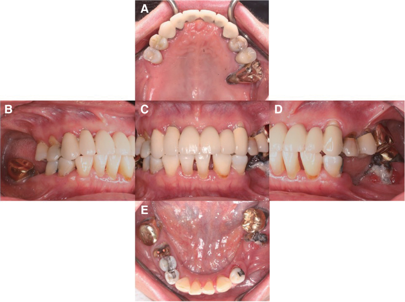
Fig. 1 Intraoral photograph. (A) Maxillary occlusal view, (B) Right lateral view, (C) Frontal view, (D) Left lateral view, (E) Mandibular occlusal view.

Fig. 2 (A) Frontal image of model after tooth removal, (B) Frontal image after Diagnostic wax up, (C) Maxilla superimposition image, (D) Mandible superimposition image, (E) Maxillary tooth STL file, (F) Mandibular tooth STL file.

Fig. 3 (A) Diagnostic wax up occlusal view of maxilla, (B) Diagnostic wax up left lateral view, (C) Diagnostic wax up occlusal view of mandible, (D) Tooth alignment occlusal view of maxilla, (E) Tooth alignment left lateral view, (F) Tooth alignment occlusal view of mandible, (G) Interim denture occlusal view of maxilla, (H) Interim denture left lateral view, (I) Interim denture occlusal view of mandible.
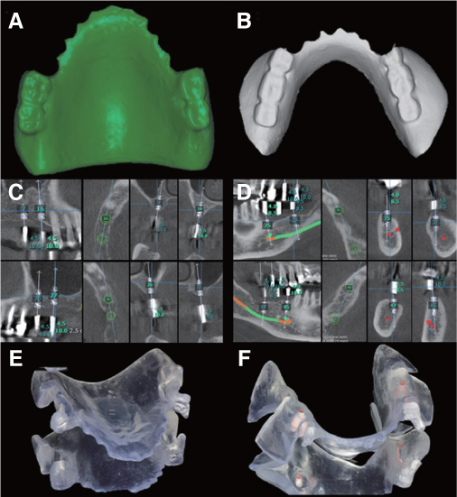
Fig. 4 (A) Image of Maxillary interim denture, (B) Mandibular interim denture, (C) Maxilla implant planning, (D) Mandible implant planning, (E) Maxilla implant surgical template, (F) Mandible implant surgical template.
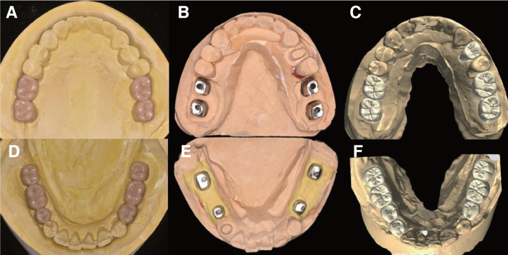
Fig. 5 (A) Diagnostic wax up occlusal view of maxilla, (B) Custom abutment occlusal view of maxilla, (C) Zirconia crown design of Maxilla, (D) Diagnostic wax up occlusal view of mandible, (E) Custom abutment occlusal view of mandible, (F) Zirconia crown design of mandible.
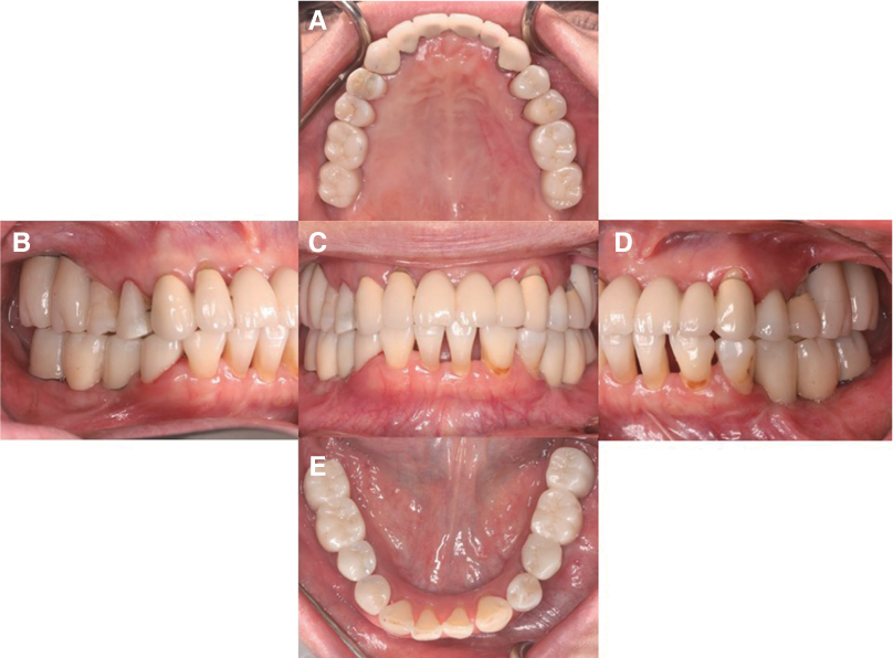
Fig. 6 Final prostheses delivery. (A) Occlusal view of maxilla, (B) Right lateral view, (C) Frontal view, (D) Left lateral view, (E) Mandibular occlusal view.
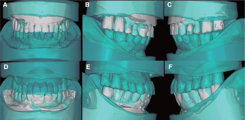
Fig. 7 Superimposition between Diagnostic wax up (sky blue) and Final prostheses (white), (A) Maxilla Frontal view, (B) Maxilla right lateral view, (C) Maxilla left lateral view, (D) Mandible Frontal view, (E) Mandible right lateral view, (F) Mandible left lateral view.
Cited by 1 articles
-
Fabrication of surveyed crown and repairing the artificial teeth for existing removable partial denture using digital technology: a case report
Ina Kim, Eunji Oh, Sang-Won Park, Hyun-Pil Lim, Kwi-dug Yun, Chan Park
J Dent Rehabil Appl Sci. 2024;40(2):82-90. doi: 10.14368/jdras.2024.40.2.82.
Reference
-
1. Davidowitz G, Kotick PG. The use of CAD/CAM in dentistry. Dent Clin North Am. 2011; 55:559–570.
Article2. Birnbaum N, Aaronson H, Stevens C, Cohen B. 3D digital scanners: A high-tech approach to more accurate dental impressions. Inside Dent. 2009; 5:70–74.3. Park JM, Koak JY, Kwon HB, Kim MJ, Kim SK, Kim SH, Yeo IS, Lim YJ, Han JS, Heo SJ. Implant-supported restoration cases fabricated from digital impression data with the help of intraoral scanner and virtual articulator. Implantology. 2017; 21:14–23.
Article4. Jung RE, Pjetursson BE, Glauser R, Zembic A, Zwahlen M, Lang NP. A systematic review of the 5-year survival and complication rates of implant-supported single crowns. Clin Oral Implants Res. 2008; 19:119–130.
Article5. Buser D, Martin W, Belser UC. Optimizing esthetics for implant restorations in the anterior maxilla: anatomic and surgical considerations. Int J Oral Maxillofac Implants. 2004; 19:43–61.6. Smeets R, Stadlinger B, Schwarz F, Beck-Broichsitter B, Jung O, Precht C, Kloss F, Gröbe A, Heiland M, Ebker T. Impact of dental implant surface modifications on osseointegration. Biomed Res Int. 2016; 2016:6285620.
Article7. Hämmerle CH, Jung RE, Feloutzis A. A systematic review of the survival of implants in bone sites augmented with barrier membranes (guided bone regeneration) in partially edentulous patients. J Clin Periodontol. 2002; 29:226–231.
Article8. Khraisat A, Hashimoto A, Nomura S, Miyakawa O. Effect of lateral cyclic loading on abutment screw loosening of an external hexagon implant system. J Prosthet Dent. 2004; 91:326–334.
Article9. Garber DA, Belser UC. Restoration-driven implant placement with restoration-generated site development. Compend Contin Educ Dent. 1995; 16:796.10. BouSerhal C, Jacobs R, Quirynen M, van Steenberghe D. Imaging technique selection for the preoperative planning of oral implants: a review of the literature. Clin Implant Dent Relat Res. 2002; 4:156–172.
Article11. Belser UC, Mericske-Stern R, Bernard JP, Taylor TD. Prosthetic management of the partially dentate patient with fixed implant restorations. Clin Oral Implants Res. 2000; 11:126–145.
Article12. Furuse AY, Baratto SS, Spina DR, Correr GM, da Cunha LF, Gonzaga CC. Planning extensive esthetic restorations for anterior teeth: use of waxed-up study casts and composite resin mock-ups. Gen Dent. 2016; 64:e6–e9.13. Beuer F, Schweiger J, Edelhoff D. Digital dentistry: an overview of recent developments for CAD/CAM generated restorations. Br Dent J. 2008; 204:505–511.
Article14. Park JM, Kim J, Shim JS. Review of computerassisted implant surgeries: Navigation surgery system vs. Computerguided implant template vs. robot. Implantology. 2018; 22:56–65.15. Schulter CW. Custom-made denture teeth and denture base material for removable partial dentures. J Prosthet Dent. 1985; 53:271–276.
Article16. Reis KR, Bonfante G, Pegoraro LF, Conti PC, Oliveira PC, Kaizer OB. In vitro wear resistance of three types of polymethyl methacrylate denture teeth. J Appl Oral Sci. 2008; 16:176–180.
Article17. D'Souza KM, Aras MA. Types of implant surgical guides in dentistry: a review. J Oral Implantol. 2012; 38:643–652.
- Full Text Links
-
- Actions
-
Cited
- CITED
-
- Close
- Share
-
- Similar articles
-
- Full mouth rehabilitation of an edentulous patient using maxillary complete denture and mandibular implant supported fixed prostheses: a case report
- Digital interim immediate denture fabrication and implant-supported removable partial denture fabrication after multiple teeth extraction in patient with chronic periodontitis: a case report
- Complete mouth rehabilitation with fixed implant-supported prosthesis using temporary denture and dental CAD-CAM
- Implant Surgery for Fixed Implant-supported Prostheses in the Edentulous Mandible: A Case Report
- Full mouth rehabilitation utilizing computer guided implant surgery and CAD/CAM
