Minimally Invasive Surgery for Hallux Valgus Deformity Using Intramedullary Low Profile Plate Fixation: A Case Report
- Affiliations
-
- 1Department of Orthopedic Surgery, Inje University Ilsan Paik Hospital, Inje University College of Medicine, Goyang, Korea. osddr8151@paik.ac.kr
- KMID: 2458016
- DOI: http://doi.org/10.14193/jkfas.2019.23.3.135
Abstract
- According to a recent systemic review, hallux valgus deformity has a prevalence rate of about 23% among adults aged 18 to 65 years. To date, more than 100 operative methods have been reported for the correction of hallux valgus deformity. For young female with mild to moderate hallux valgus deformity, minimally invasive surgery can be considered for aesthetic demands. Here, we report a case of a young female patient with mild hallux valgus deformity treated by minimally invasive surgery using intramedullary low profile plate fixation. This can be the favorable method for secure fixation of the osteotomy site and prevention of medial skin irritation symptoms derived from a sharp osteotomy margin.
MeSH Terms
Figure
-
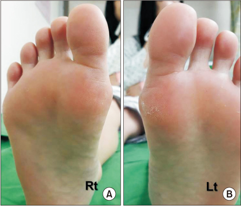
Figure 1 Bilateral hallux valgus deformity with mild plantar keratosis was noted. (A) Right foot. (B) Left Foot.
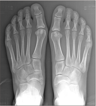
Figure 2 On the weight bearing anteroposterior foot radiograph, bilateral mild hallux valgus deformity was seen.
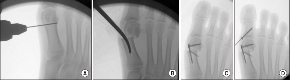
Figure 3 (A) Through the 8 mm medial incision, transverse osteotomy on the metatarsal neck was performed with oscillating saw. (B) By inserting the curved Kelly forcep to the medullary space, distal fragment was spontaneously translated to the lateral side. (C) Then Low profile locking plate was put intramedullary and locking screws were fixed to secure the osteotomy site. (D) Additional Akin procedure was done with 3.0 cannulated screw fixation.

Figure 4 Postoperative scar formation at 1-year follow-up.
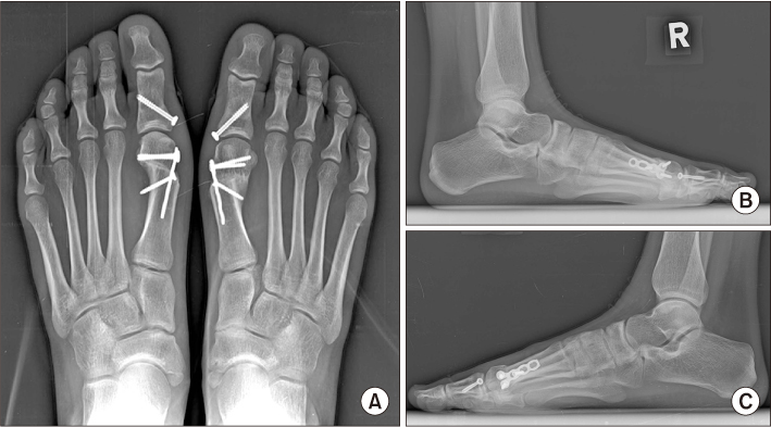
Figure 5 At postoperative 1 year, weight bearing foot anteroposterior radiograph (A) and right (B) and left (C) lateral radiographs show the adequate correction of the deformity.
Cited by 1 articles
-
Minimally Invasive Proximal Transverse Metatarsal Osteotomy Followed by Intramedullary Plate Fixation for Hallux Valgus Deformity: A Case Report
Jong Hun Kim, Jin Soo Suh, Jun Young Choi
J Korean Foot Ankle Soc. 2021;25(3):141-144. doi: 10.14193/jkfas.2021.25.3.141.
Reference
-
1. Robinson AH, Limbers JP. Modern concepts in the treatment of hallux valgus. J Bone Joint Surg Br. 2005; 87:1038–1045. DOI: 10.1302/0301-620X.87B8.16467.
Article2. Bosch P, Markowski H, Rannicher V. [Technik und erste ergebnisse der subkutanen distalen metatarsale, I osteotomie]. Orthop Prax. 1990; 26:51–56. German.3. Faour-Martín O, Martín-Ferrero MA, Valverde García JA, Vega-Castrillo A, de la Red-Gallego MA. Long-term results of the retrocapital metatarsal percutaneous osteotomy for hallux valgus. Int Orthop. 2013; 37:1799–1803. DOI: 10.1007/s00264-013-1934-1.
Article4. Magnan B, Pezzè L, Rossi N, Bartolozzi P. Percutaneous distal metatarsal osteotomy for correction of hallux valgus. J Bone Joint Surg Am. 2005; 87:1191–1199. DOI: 10.2106/JBJS.D.02280.
Article5. Kadakia AR, Smerek JP, Myerson MS. Radiographic results after percutaneous distal metatarsal osteotomy for correction of hallux valgus deformity. Foot Ankle Int. 2007; 28:355–360. DOI: 10.3113/FAI.2007.0355.
Article6. Huang PJ, Lin YC, Fu YC, Yang YH, Cheng YM. Radiographic evaluation of minimally invasive distal metatarsal osteotomy for hallux valgus. Foot Ankle Int. 2011; 32:S503–S507. DOI: 10.3113/FAI.2011.0503.
Article7. Choi JY, Ahn HC, Kim SH, Lee SY, Suh JS. Minimally invasive surgery for young female patients with mild-to-moderate juvenile hallux valgus deformity. Foot Ankle Surg. 2019; 25:316–322. DOI: 10.1016/j.fas.2017.12.006.
Article8. Giannini S, Cavallo M, Faldini C, Luciani D, Vannini F. The SERI distal metatarsal osteotomy and Scarf osteotomy provide similar correction of hallux valgus. Clin Orthop Relat Res. 2013; 471:2305–2311. DOI: 10.1007/s11999-013-2912-z.
Article9. Enan A, Abo-Hegy M, Seif H. Early results of distal metatarsal osteotomy through minimally invasive approach for mild-to-moderate hallux valgus. Acta Orthop Belg. 2010; 76:526–535.10. Brogan K, Voller T, Gee C, Borbely T, Palmer S. Third-generation minimally invasive correction of hallux valgus: technique and early outcomes. Int Orthop. 2014; 38:2115–2121. DOI: 10.1007/s00264-014-2500-1.
Article
- Full Text Links
-
- Actions
-
Cited
- CITED
-
- Close
- Share
-
- Similar articles
-
- Minimally Invasive Proximal Transverse Metatarsal Osteotomy Followed by Intramedullary Plate Fixation for Hallux Valgus Deformity: A Case Report
- Minimally Invasive Surgery with Tenorrhaphy for Postoperative Hallux Varus Deformity Combined with Flexor Hallucis Longus Rupture after Hallux Valgus Correction: A Case Report
- Minimally Invasive Distal Transverse Metatarsal Osteotomy – Akin Osteotomy (MITA) for Recurrent Hallux Valgus: A Report of Four Cases
- The Clinical Results of the Proximal Opening Wedge Osteotomy Using a Low Profile Plate in Hallux Valgus: Comparison with Proximal Chevron Osteotomy Fixed with K-wires
- Bioabsorbable Screws Used in Hallux Valgus Treatment Using Proximal Chevron Osteotomy
