Minimally Invasive Surgery with Tenorrhaphy for Postoperative Hallux Varus Deformity Combined with Flexor Hallucis Longus Rupture after Hallux Valgus Correction: A Case Report
- Affiliations
-
- 1Department of Orthopaedic Surgery, Ilsan Paik Hospital, Inje University College of Medicine, Goyang, Korea
- KMID: 2502918
- DOI: http://doi.org/10.14193/jkfas.2020.24.2.102
Abstract
- A postoperative hallux varus deformity is a dreaded complication of hallux valgus surgery. Several surgical options have been introduced to overcome this problem. This paper reports an uncommon case of a 68-year-old female patient who presented with a postoperative hallux varus deformity combined with a rupture of the flexor hallucis longus (FHL) tendon. She was treated successfully by a minimally invasive correctional osteotomy with open tenorrhaphy. With experience in treating this complicated case, it was noted that FHL could be transected during the trans-articular adductor tenotomy. Hence, extra caution is needed when the degree of hallux valgus deformity is excessive. To the best of the author’s knowledge, correctional valgization osteotomy for a postoperative hallux varus deformity in a minimally invasive manner has not been reported. This case report is expected to benefit surgeons and their patients with severe hallux valgus deformity.
Keyword
Figure
-
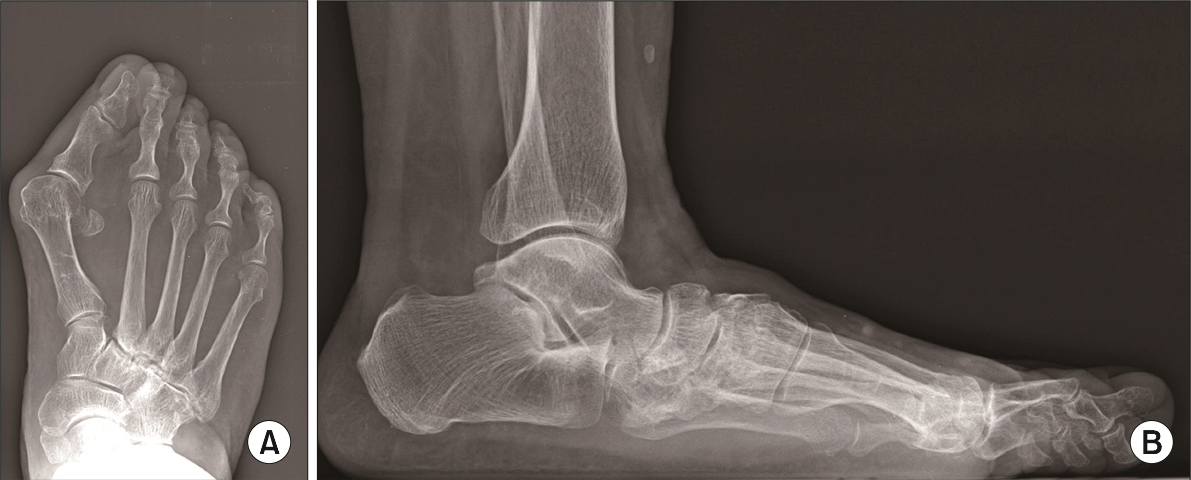
Figure. 1 On the weight bearing anteroposterior (A) and lateral (B) foot radiographs, severe hallux valgus with pes planus deformity was seen. Preoperative hallux valgus angle and 1st to 2nd intermetatarsal angle were 48.0° and 24.5°, respectively.

Figure. 2 Immediate postoperative radiograph (A) shows the enough correction of prior deformity while hallux varus deformity was occurred at 6 weeks (B). Degree of varus deformity became greater with great toe flexion limitation at postoperative 3 months (C) and it persisted after the removal of K-wires (D). Patient complained the discomfort while she worn the shoe (E).
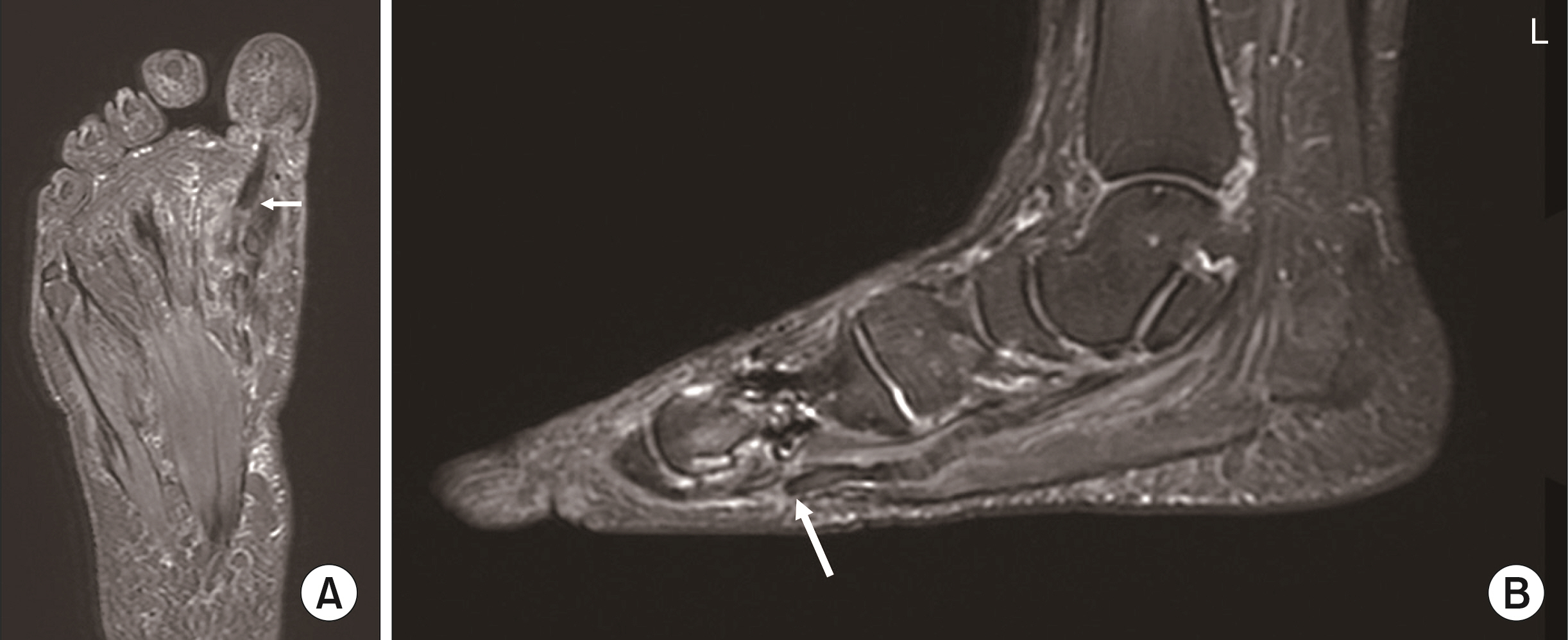
Figure. 3 Coronal (A) and sagittal (B) magnetic resonance images show the complete rupture of flexor hallucis longus tendon around the 1st metatarso-phalangeal joint (arrows).
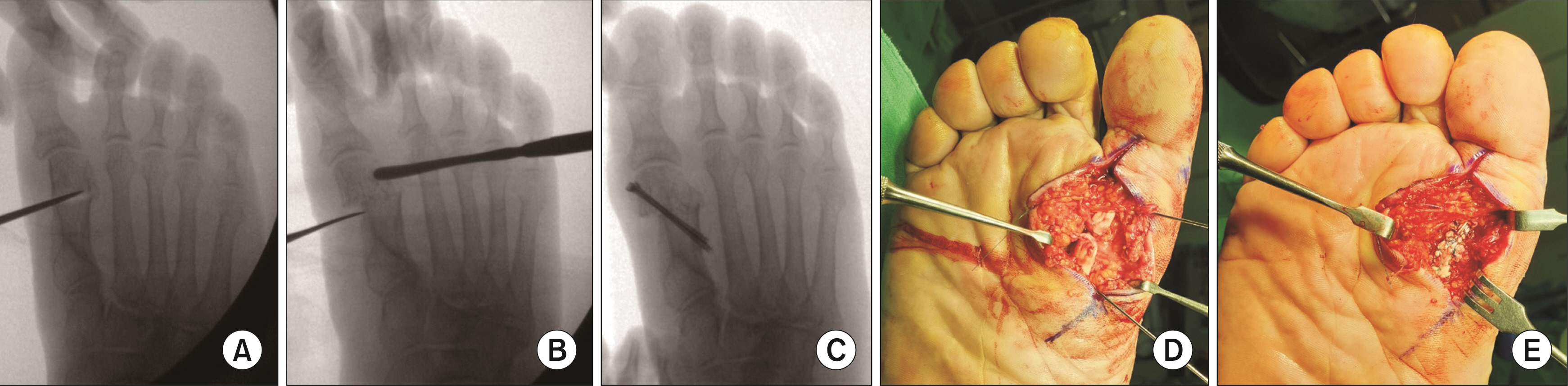
Figure. 4 (A) Through the 1 cm medial incision, transverse osteotomy on the metatarsal neck was performed with a conventional oscillating saw. (B) Distal fragment was pushed medially with a freer elevator. (C) Then two 3.0 mm cannulated screws were fixed through the osteotomy site. We could note the rupture of flexor hallucis longus tendon through another incision from the sole (D) and repair it by Becker’s method (E).
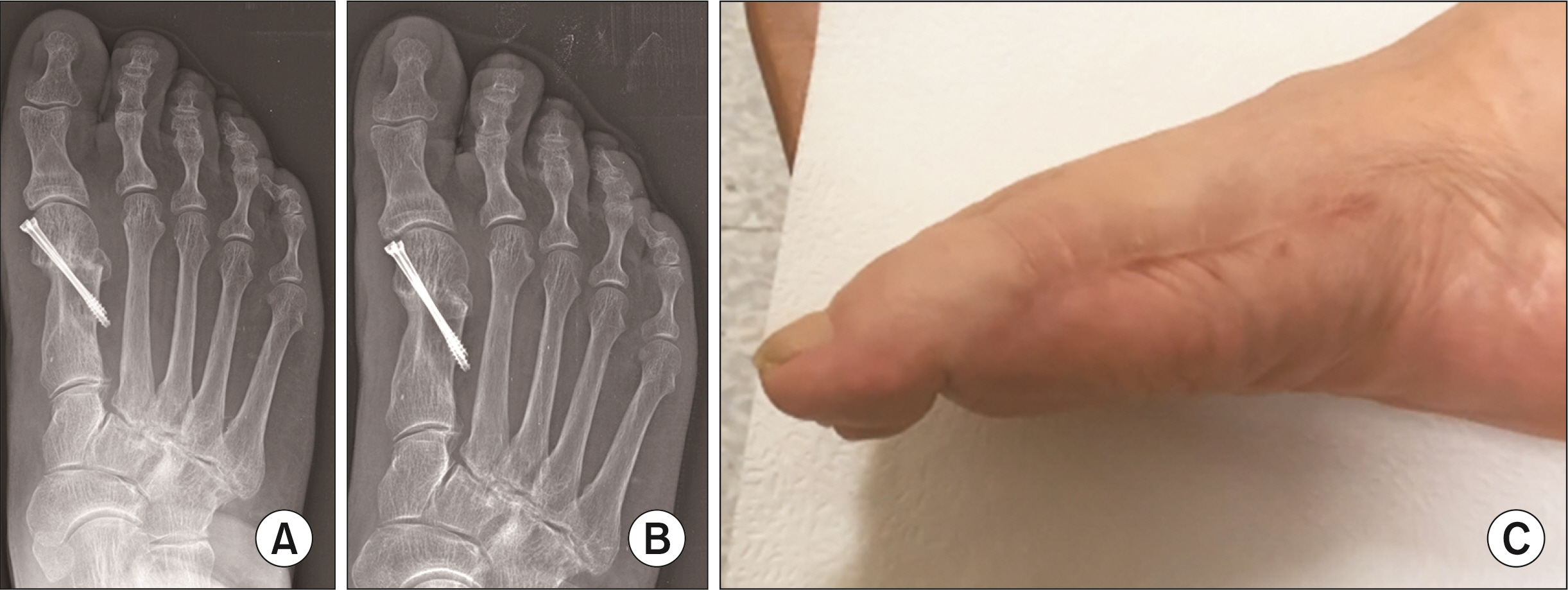
Figure. 5 Postoperative radiographs at 3 months (A) and 1 year (B) after the revision show the maintenance of correction. (C) The maximal active plantar flexion of the interphalangeal joint was 25°.
Reference
-
1. Robinson AH, Limbers JP. 2005; Modern concepts in the treatment of hallux valgus. J Bone Joint Surg Br. 87:1038–45. doi: 10.1302/0301-620X.87B8.16467. DOI: 10.1302/0301-620X.87B8.16467. PMID: 16049235.
Article2. Bösch P, Markowski H, Rannicher V. 1990; [Technik und erste ergebnisse der subkutanen distalen metatarsale-I-osteotomie]. Orthop Praxis. 26:51–6.3. Faour-Martín O, Martín-Ferrero MA, Valverde García JA, Vega-Castrillo A, de la Red-Gallego MA. 2013; Long-term results of the retrocapital metatarsal percutaneous osteotomy for hallux valgus. Int Orthop. 37:1799–803. doi: 10.1007/s00264-013-1934-1. DOI: 10.1007/s00264-013-1934-1. PMID: 23722318. PMCID: PMC3764279.
Article4. Sammarco GJ, Idusuyi OB. 2001; Complications after surgery of the hallux. Clin Orthop Relat Res. (391):59–71. doi: 10.1097/00003086-200110000-00008. DOI: 10.1097/00003086-200110000-00008. PMID: 11603690.
Article5. Schuh R, Willegger M, Holinka J, Ristl R, Windhager R, Wanivenhaus AH. 2013; Angular correction and complications of proximal first metatarsal osteotomies for hallux valgus deformity. Int Orthop. 37:1771–80. doi: 10.1007/s00264-013-2012-4. DOI: 10.1007/s00264-013-2012-4. PMID: 23884327. PMCID: PMC3764281.
Article6. Raikin SM, Miller AG, Daniel J. 2014; Recurrence of hallux valgus: a review. Foot Ankle Clin. 19:259–74. doi: 10.1016/j.fcl.2014.02.008. DOI: 10.1016/j.fcl.2014.02.008. PMID: 24878414.7. Crawford MD, Patel J, Giza E. 2014; Iatrogenic hallux varus treatment algorithm. Foot Ankle Clin. 19:371–84. doi: 10.1016/j.fcl.2014.06.004. DOI: 10.1016/j.fcl.2014.06.004. PMID: 25129350.
Article8. Lee KT, Young KW, Bae SW, Bang YS, Kim DH. 2003; Iatrogenic hallux varus deformity after hallux valgus surgery. J Korean Soc Foot Surg. 7:101–8.9. Choi KJ, Lee HS, Yoon YS, Park SS, Kim JS, Jeong JJ, et al. 2011; Distal metatarsal osteotomy for hallux varus following surgery for hallux valgus. J Bone Joint Surg Br. 93:1079–83. doi: 10.1302/0301-620X.93B8.26430. DOI: 10.1302/0301-620X.93B8.26430. PMID: 21768632.
Article10. Brogan K, Voller T, Gee C, Borbely T, Palmer S. 2014; Third-generation minimally invasive correction of hallux valgus: technique and early outcomes. Int Orthop. 38:2115–21. doi: 10.1007/s00264-014-2500-1. DOI: 10.1007/s00264-014-2500-1. PMID: 25128969.
Article11. Devos Bevernage B, Leemrijse T. 2009; Hallux varus: classification and treatment. Foot Ankle Clin. 14:51–65. doi: 10.1016/j.fcl.2008.11.007. DOI: 10.1016/j.fcl.2008.11.007. PMID: 19232992.
Article12. Brand JC Jr, Smith RW. 1991; Rupture of the flexor hallucis longus after hallux valgus surgery: case report and comments on technique for adductor release. Foot Ankle. 11:407–10. doi: 10.1177/107110079101100614. DOI: 10.1177/107110079101100614. PMID: 1894238.
Article13. Gillott E, Ray PS. 2012; Repair of iatrogenic rupture of the flexor hallucis longus tendon following an Akin osteotomy: a case report and review of literature. Foot Ankle Online J. 5:1.
- Full Text Links
-
- Actions
-
Cited
- CITED
-
- Close
- Share
-
- Similar articles
-
- Minimally Invasive Proximal Transverse Metatarsal Osteotomy Followed by Intramedullary Plate Fixation for Hallux Valgus Deformity: A Case Report
- Minimally Invasive Surgery for Hallux Valgus Deformity Using Intramedullary Low Profile Plate Fixation: A Case Report
- Complications after Surgical Correction of Hallux Valgus
- Reconstruction of Chronic Extensor Hallucis Longus Tendon Rupture Using Interposed Scar Tissue: A Case Report
- Management of Checkrein Deformity
