J Adv Prosthodont.
2019 Aug;11(4):215-222. 10.4047/jap.2019.11.4.215.
Influence of polishing systems on roughness and color change of two dental ceramics
- Affiliations
-
- 1Dental School, University of Taubaté - UNITAU, Taubaté, São Paulo, Brazil. lucascmaciel@hotmail.com
- 2Dental School, School of Higher Education of São Francisco de Assis - ESFA, Santa Teresa, EspÃrito Santo, Brazil.
- 3Dental School, University Veiga de Almeida - UVA, Rio de Janeiro, Rio de Janeiro, Brazil.
- 4Dental School, Federal University of EspÃrito Santo - UFES, Vitória, EspÃrito Santo, Brazil.
- KMID: 2455930
- DOI: http://doi.org/10.4047/jap.2019.11.4.215
Abstract
- PURPOSE
To evaluate the polishing effect on roughness and color change of pressed and layering ceramics after immersion in coffee solution.
MATERIALS AND METHODS
88 ceramic discs (1.0 mm × 10.0 mm) were manufactured - 44 nano-fluorapatite layering ceramics (IPS e.max Ceram. Group C) and 44 pressed lithium disilicate ceramic discs (IPS e. max Press - Group P). Each group was divided into 4 subgroups according to surface treatments: (G) Glaze, (S) Shofu polishing system (Shofu Inc.), (E) Edenta AG polishing System, (KG) 30-µm diamond granulation tip. Surface roughness (Ra) and color change (ΔE) measurings after the surface treatments were performed, before and 12 days after the immersion in coffee solution. A samples' qualitative analysis was conducted with a scanning electron microscopy (SEM). Data were statistically-treated with one-way-ANOVA and Duncan's tests, apart from paired t-test and Pearson's correlation test (α=5%).
RESULTS
The decrescent order, both for surface roughness (Ra) and ΔE for both ceramics were: KG > E > S > G (P<.05). With exception for PG and CG subgroups, which did not present statistical difference between them, all other pressed ceramics subgroups presented smaller Ra values and greater ΔE values than the layering ceramics subgroups (P<.05).
CONCLUSION
Although mechanical polishing systems presented intermediate Ra values, their colors were considered clinically acceptable. There is a strong correlation between the surface roughness and the color change of tested ceramics.
MeSH Terms
Figure
-
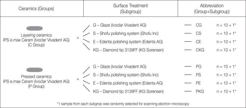
Fig. 1 Experimental sketch. Ceramics and surface treatments applied in this study.
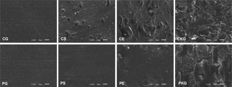
Fig. 2 Scanning electron microscopy (× 1,000) of layering ceramics (C Group) and pressed ceramics (P Group) after different surface treatments: (G) Glaze, (S) Shofu Inc polishing system, (E) Edenta AG polishing system and (KG) 30 µm diamond tip.
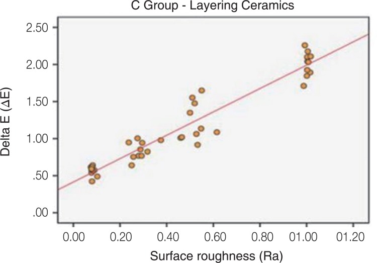
Fig. 3 Pearson correlation analysis. Graph of dispersal between the surface roughness (Ra) and the color change (ΔE) in Group C.
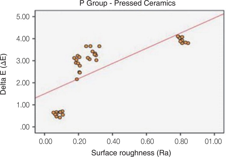
Fig. 4 Pearson correlation analysis. Graph of dispersal between the surface roughness (Ra) and the color change (ΔE) in Group P.
Reference
-
1. Höland W, Rheinberger V, Apel E, Ritzberger C, Rothbrust F, Kappert H, Krumeich F, Nesper R. Future perspectives of biomaterials for dental restoration. J European Ceram Soc. 2009; 29:1291–1297.
Article2. Bayne SC, Ferracane JL, Marshall GW, Marshall SJ, van Noort R. The evolution of dental materials over the past century: Silver and gold to tooth color and beyond. J Dent Res. 2019; 98:257–265. PMID: 30784370.
Article3. de Kok P, Pereira GKR, Fraga S, de Jager N, Venturini AB, Kleverlaan CJ. The effect of internal roughness and bonding on the fracture resistance and structural reliability of lithium disilicate ceramic. Dent Mater. 2017; 33:1416–1425. PMID: 29032826.
Article4. Dalkiz M, Sipahi C, Beydemir B. Effects of six surface treatment methods on the surface roughness of a low-fusing and an ultra low-fusing feldspathic ceramic material. J Prosthodont. 2009; 18:217–222. PMID: 19141045.
Article5. Kelly JR, Benetti P. Ceramic materials in dentistry: historical evolution and current practice. Aust Dent J. 2011; 56:84–96. PMID: 21564119.
Article6. Anusavice KJ, Shen C, Rawls HR. Phillips' science of dental materials. 12th ed. St. Louis: Elsevier/Saunders;2012. p. 418–473.7. Wright MD, Masri R, Driscoll CF, Romberg E, Thompson GA, Runyan DA. Comparison of three systems for the polishing of an ultra-low fusing dental porcelain. J Prosthet Dent. 2004; 92:486–490. PMID: 15523338.
Article8. Chang CW, Waddell JN, Lyons KM, Swain MV. Cracking of porcelain surfaces arising from abrasive grinding with a dental air turbine. J Prosthodont. 2011; 20:613–620. PMID: 22017480.
Article9. al-Wahadni A, Martin DM. Glazing and finishing dental porcelain: a literature review. J Can Dent Assoc. 1998; 64:580–583. PMID: 9785688.10. Rashid H. The effect of surface roughness on ceramics used in dentistry: A review of literature. Eur J Dent. 2014; 8:571–579. PMID: 25512743.
Article11. Yilmaz C, Korkmaz T, Demirköprülü H, Ergün G, Ozkan Y. Color stability of glazed and polished dental porcelains. J Prosthodont. 2008; 17:20–24. PMID: 17971115.12. Vichi A, Fonzar RF, Goracci C, Carrabba M, Ferrari M. Effect of finishing and polishing on roughness and gloss of lithium disilicate and lithium silicate zirconia reinforced glass ceramic for CAD/CAM systems. Oper Dent. 2018; 43:90–100. PMID: 29284101.
Article13. Yuzugullu B, Celik C, Erkut S, Ozcelik TB. The effects of extraoral porcelain polishing sequences on surface roughness and color of feldspathic porcelain. Int J Prosthodont. 2009; 22:472–475. PMID: 20095196.14. Sarikaya I, Güler AU. Effects of different polishing techniques on the surface roughness of dental porcelains. J Appl Oral Sci. 2010; 18:10–16. PMID: 20379676.
Article15. Al-Shammery HA, Bubb NL, Youngson CC, Fasbinder DJ, Wood DJ. The use of confocal microscopy to assess surface roughness of two milled CAD-CAM ceramics following two polishing techniques. Dent Mater. 2007; 23:736–741. PMID: 16914192.
Article16. Mohammadibassir M, Rezvani MB, Golzari H, Moravej Salehi E, Fahimi MA, Kharazi Fard MJ. Effect of two polishing systems on surface roughness, topography, and flexural strength of a monolithic lithium disilicate ceramic. J Prosthodont. 2019; 28:e172–e180. PMID: 28273681.
Article17. Turgut S, Bagis B. Colour stability of laminate veneers: an in vitro study. J Dent. 2011; 39:e57–e64. PMID: 22101122.
Article18. Lee YK, Yu B, Lim JI, Lim HN. Perceived color shift of a shade guide according to the change of illuminant. J Prosthet Dent. 2011; 105:91–99. PMID: 21262406.
Article19. Kelly JR. Ceramics in restorative and prosthetic dentistry. Annual Rev Mater Sci. 1997; 27:443–468.
Article20. Sarikaya I, Güler AU. Effects of different surface treatments on the color stability of various dental porcelains. J Dent Sci. 2011; 6:65–71.
Article21. Motro PF, Kursoglu P, Kazazoglu E. Effects of different surface treatments on stainability of ceramics. J Prosthet Dent. 2012; 108:231–237. PMID: 23031729.
Article22. Kursoglu P, Karagoz Motro PF, Kazazoglu E. Correlation of surface texture with the stainability of ceramics. J Prosthet Dent. 2014; 112:306–313. PMID: 24484857.
Article23. Sarıkaya I, Yerliyurt K, Hayran Y. Effect of surface finishing on the colour stability and translucency of dental ceramics. BMC Oral Health. 2018; 18:40. PMID: 29534712.
Article24. Sarac D, Sarac YS, Yuzbasioglu E, Bal S. The effects of porcelain polishing systems on the color and surface texture of feldspathic porcelain. J Prosthet Dent. 2006; 96:122–128. PMID: 16911889.
Article25. Camacho GB, Vinha D, Panzeri H, Nonaka T, Gonçalves M. Surface roughness of a dental ceramic after polishing with different vehicles and diamond pastes. Braz Dent J. 2006; 17:191–194. PMID: 17262123.
Article26. Saraç D, Turk T, Elekdag-Turk S, Saraç YS. Comparison of 3 polishing techniques for 2 all-ceramic materials. Int J Prosthodont. 2007; 20:465–468. PMID: 17944333.27. Boaventura JM, Nishida R, Elossais AA, Lima DM, Reis JM, Campos EA, de Andrade MF. Effect finishing and polishing procedures on the surface roughness of IPS Empress 2 ceramic. Acta Odontol Scand. 2013; 71:438–443. PMID: 22724660.
Article28. Carrabba M, Vichi A, Vultaggio G, Pallari S, Paravina R, Ferrari M. Effect of finishing and polishing on the surface roughness and gloss of feldspathic ceramic for chairside CAD/CAM systems. Oper Dent. 2017; 42:175–184. PMID: 27723423.
Article29. Ertan AA, Sahin E. Colour stability of low fusing porcelains: an in vitro study. J Oral Rehabil. 2005; 32:358–361. PMID: 15842245.
Article30. Samra AP, Pereira SK, Delgado LC, Borges CP. Color stability evaluation of aesthetic restorative materials. Braz Oral Res. 2008; 22:205–210. PMID: 18949304.
Article31. Niu E, Agustin M, Douglas RD. Color match of machinable lithium disilicate ceramics: effects of foundation restoration. J Prosthet Dent. 2013; 110:501–509. PMID: 24169080.
Article32. Akar GC, Pekkan G, Çal E, Eskitaşçıoğlu G, Özcan M. Effects of surface-finishing protocols on the roughness, color change, and translucency of different ceramic systems. J Prosthet Dent. 2014; 112:314–321. PMID: 24513427.33. Niu E, Agustin M, Douglas RD. Color match of machinable lithium disilicate ceramics: Effects of cement color and thickness. J Prosthet Dent. 2014; 111:42–50. PMID: 24210729.
Article34. Alp G, Subasi MG, Johnston WM, Yilmaz B. Effect of surface treatments and coffee thermocycling on the color and translucency of CAD-CAM monolithic glass-ceramic. J Prosthet Dent. 2018; 120:263–268. PMID: 29551378.
Article35. Atay A, Karayazgan B, Ozkan Y, Akyil MS. Effect of colored beverages on the color stability of feldspathic porcelain subjected to various surface treatments. Quintessence Int. 2009; 40:e41–e48. PMID: 19626223.36. Sagsoz O, Demirci T, Demirci G, Sagsoz NP, Yildiz M. The effects of different polishing techniques on the staining resistance of CAD/CAM resin-ceramics. J Adv Prosthodont. 2016; 8:417–422. PMID: 28018558.
Article37. Guler AU, Yilmaz F, Kulunk T, Guler E, Kurt S. Effects of different drinks on stainability of resin composite provisional restorative materials. J Prosthet Dent. 2005; 94:118–124. PMID: 16046965.
Article38. Raimondo RL Jr, Richardson JT, Wiedner B. Polished versus autoglazed dental porcelain. J Prosthet Dent. 1990; 64:553–557. PMID: 2090814.
Article39. Patterson CJ, McLundie AC, Stirrups DR, Taylor WG. Efficacy of a porcelain refinishing system in restoring surface finish after grinding with fine and extra-fine diamond burs. J Prosthet Dent. 1992; 68:402–406. PMID: 1331431.
Article40. Goldstein GR, Barnhard BR, Penugonda B. Profilometer, SEM, and visual assessment of porcelain polishing methods. J Prosthet Dent. 1991; 65:627–634. PMID: 2051383.
Article41. Manjuran NG, Sreelal T. An in vitro study to identify a ceramic polishing protocol effecting smoothness superior to glazed surface. J Indian Prosthodont Soc. 2014; 14:219–227. PMID: 25183905.
Article42. Steiner R, Beier US, Heiss-Kisielewsky I, Engelmeier R, Dumfahrt H, Dhima M. Adjusting dental ceramics: An in vitro evaluation of the ability of various ceramic polishing kits to mimic glazed dental ceramic surface. J Prosthet Dent. 2015; 113:616–622. PMID: 25794914.43. Fuzzi M, Zaccheroni Z, Vallania G. Scanning electron microscopy and profilometer evaluation of glazed and polished dental porcelain. Int J Prosthodont. 1996; 9:452–458. PMID: 9108746.44. Bottino MC, Valandro LF, Kantorski KZ, Bressiani JC, Bottino MA. Polishing methods of an alumina-reinforced feldspar ceramic. Braz Dent J. 2006; 17:285–289. PMID: 17262140.
Article45. Seghi RR, Hewlett ER, Kim J. Visual and instrumental colorimetric assessments of small color differences on translucent dental porcelain. J Dent Res. 1989; 68:1760–1764. PMID: 2600257.
Article46. Gupta R, Parkash H, Shah N, Jain V. A spectrophotometric evaluation of color changes of various tooth colored veneering materials after exposure to commonly consumed beverages. J Indian Prosthodont Soc. 2005; 5:72–78.
Article
- Full Text Links
-
- Actions
-
Cited
- CITED
-
- Close
- Share
-
- Similar articles
-
- The effect of repeated firings on the color change and surface roughness of dental ceramics
- The effect of various polishing systems on surface roughness and phase transformation of monolithic zirconia
- Comparison of Color Stability and Surface Roughness of 3D Printing Resin by Polishing Methods
- Surface roughness and color stability of various composite resins
- The evaluation of surface roughness and polishing time between polishing systems
