Clin Endosc.
2019 Jul;52(4):360-364. 10.5946/ce.2018.160.
Diagnostic Efficacy of Endoscopic Ultrasound Elastography in Differentiating Solid Pancreatic Lesions: A Single-Center Experience
- Affiliations
-
- 1Department of Gastroenterology and Hepatology, Mansoura Specialized Medical Hospital, Mansoura University, Mansoura, Egypt. a.tonbary@gmail.com
- 2Department of Anesthesia and Intensive Care, Mansoura University, Mansoura, Egypt.
- KMID: 2455633
- DOI: http://doi.org/10.5946/ce.2018.160
Abstract
- BACKGROUND/AIMS
Endoscopic ultrasound (EUS) has a limited ability to determine the nature of solid pancreatic lesions (SPLs). Most recent ultrasound processors are provided with elastography software, which allows quantification of the tissue hardness. The aim of this study is to evaluate the effectiveness of the elasticity score (ES) and strain ratio (SR) in the differentiation of benign pancreatic lesions from malignant pancreatic lesions.
METHODS
The study had a retrospective design; it included 97 patients with SPLs and 19 patients with inflammatory lesions. The ES and SR were determined during the examination; finally, EUS-guided fine needle aspiration was performed.
RESULTS
In this 2-year study, 116 patients were enrolled (97 with malignant lesions and 19 with benign lesions). There were 69 men and 47 women. Their median age was 55.9 years. A cut-off point was detected at SR of 7.75 with a specificity of 99.9%, sensitivity of 90.7%, positive predictive value (PPV) of 99.9%, negative predictive value (NPV) of 67.9%, and accuracy of 92.2%. After adding the ES to the SR, the cut-off point at 7.75 resulted in a specificity of 94.6%, sensitivity of 99%, PPV of 98%, NPV of 98.5%, and accuracy of 97%.
CONCLUSIONS
The use of the ES combined with the SR increases the accuracy of differentiation between benign and malignant SPLs and is an effective method for the evaluation of pancreatic masses.
MeSH Terms
Figure
-
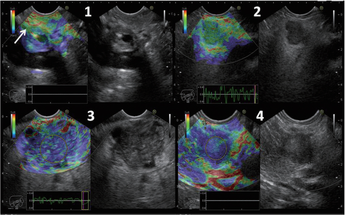
Fig. 1. Elasticity score (ES) 1 was given to homogeneous green area and indicated normal tissue (arrow). ES 2 was given to heterogeneous green area predominant and indicated fibrosis or inflammation. ES 3 was given to heterogeneous blue predominant and indicated indeterminate for malignancy. ES 4 was given to homogeneous blue and indicated malignant lesions. The strain ratio was calculated in ES 3 and 4.
Reference
-
1. Chantarojanasiri T, Kongkam P. Endoscopic ultrasound elastography for solid pancreatic lesions. World J Gastrointest Endosc. 2017; 9:506–513.
Article2. Deprez PH. EUS elastography: is it replacing or supplementing tissue acquisition? Gastrointest Endosc. 2013; 77:590–592.
Article3. Arcidiacono PG. Endoscopic ultrasound elastography. Gastroenterol Hepatol (N Y). 2012; 8:48–67.4. Iglesias-Garcia J, Larino-Noia J, Abdulkader I, Forteza J, Dominguez-Munoz JE. Quantitative endoscopic ultrasound elastography: an accurate method for the differentiation of solid pancreatic masses. Gastroenterology. 2010; 139:1172–1180.
Article5. Lee TH, Cho YD, Cha SW, et al. Endoscopic ultrasound elastography for the pancreas in Korea: a preliminary single center study. Clin Endosc. 2013; 46:172–177.
Article6. Yamabe A, Irisawa A, Bhutani MS, et al. Efforts to improve the diagnostic accuracy of endoscopic ultrasound-guided fine-needle aspiration for pancreatic tumors. Endosc Ultrasound. 2016; 5:225–232.
Article7. Eloubeidi MA, Tamhane A, Varadarajulu S, Wilcox CM. Frequency of major complications after EUS-guided FNA of solid pancreatic masses: a prospective evaluation. Gastrointest Endosc. 2006; 63:622–629.
Article8. Dawwas MF, Taha H, Leeds JS, Nayar MK, Oppong KW. Diagnostic accuracy of quantitative EUS elastography for discriminating malignant from benign solid pancreatic masses: a prospective, single-center study. Gastrointest Endosc. 2012; 76:953–961.
Article9. Giovannini M. Endoscopic ultrasound elastography. Pancreatology. 2011; 11 Suppl 2:34–39.
Article10. Giovannini M, Thomas B, Erwan B, et al. Endoscopic ultrasound elastography for evaluation of lymph nodes and pancreatic masses: a multicenter study. World J Gastroenterol. 2009; 15:1587–1593.
Article11. Okasha H, Elkholy S, El-Sayed R, et al. Real time endoscopic ultrasound elastography and strain ratio in the diagnosis of solid pancreatic lesions. World J Gastroenterol. 2017; 23:5962–5968.
Article12. Kongkam P, Lakananurak N, Navicharern P, et al. Combination of EUSFNA and elastography (strain ratio) to exclude malignant solid pancreatic lesions: a prospective single-blinded study. J Gastroenterol Hepatol. 2015; 30:1683–1689.
Article13. Iglesias-Garcia J, Lindkvist B, Lariño-Noia J, Domínguez-Muñoz JE. Endoscopic ultrasound elastography. Endosc Ultrasound. 2012; 1:8–16.
Article
- Full Text Links
-
- Actions
-
Cited
- CITED
-
- Close
- Share
-
- Similar articles
-
- Role of contrast-enhanced harmonic endoscopic ultrasonography (EUS) and EUS elastography in pancreatic lesions
- Elastography of the Pancreas, Current View
- Clinical Role of Contrast-Enhanced Harmonic Endoscopic Ultrasound in Differentiating Solid Lesions of the Pancreas: A Single-Center Experience in Korea
- How to optimize the diagnostic yield of endoscopic ultrasoundguided fine-needle sampling in solid pancreatic lesions from a technical perspective
- Clinical Efficacy of Contrast-Enhanced Endoscopic Ultrasound in the Pancreatic Solid Lesions
