Left Upper Lobar Agenesis Associated with Tracheal Trifurcation: A Case Report
- Affiliations
-
- 1Department of Radiology, Eulji Hospital, Eulji University School of Medicine, Seoul, Korea. jjblue@eulji.ac.kr
- KMID: 2454033
- DOI: http://doi.org/10.3348/jksr.2019.80.3.543
Abstract
- Lobar agenesis is a rare congenital anomaly that is characterized by the absence of the lobar pulmonary artery, pulmonary vein, bronchi, and parenchyma. We encountered a unique case of a young male patient with agenesis of the left upper lobe with tracheal trifurcation into three bronchi, all arising at the carinal level. Complex tracheobronchial anatomy was explicitly demonstrated by three-dimensional CT reconstruction and virtual bronchoscopy. Left upper lobar agenesis associated with tracheal trifurcation is an extremely rare anomaly that, to the best of our knowledge, has not been previously reported.
MeSH Terms
Figure
-
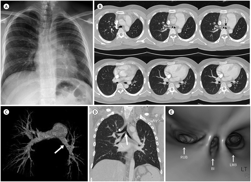
Fig. 1 A 22-year-old man exhibited agenesis of the left upper lobe, combined with tracheal trifurcation, on CT. A. Chest X-ray shows volume loss on the left side with tracheal and mediastinal shift toward the left side, as well as slight compensatory hypertrophy of the right lung field. Note the caudally displaced left hilum. B. Chest CT (lung window) axial view at subcarinal level reveals the left main bronchus (arrow), which continues as the left lower lobe bronchus with mild mediastinal shift toward the ipsilateral side, with compensatory hyperinflation of the right lung field. Note the absence of the left upper lobe. C. Surface shade display three-dimensional CT-reconstructed image reveals a hypoplastic left pulmonary (arrow), which continues as the left lower lobe pulmonary artery without any upper lobe branch. D. Chest CT (lung window) coronal view demonstrates division of the trachea into three bronchi. The right upper lobar bronchus, bronchus intermedius, and left main bronchus originate from the carina. There is relative narrowing of the proximal portion of the bronchus intermedius (arrow). E. Virtual bronchoscopic image reveals three origins of the RUB, BI, and LMB (arrows). BI = bronchus intermedius, LMB = left main bronchus, RUB = right upper lobar bronchus
Reference
-
1. Mardini MK, Nyhan WL. Agenesis of the lung. Report of four patients with unusual anomalies. Chest. 1985; 87:522–552.2. Rivera C, Gardenhire DS. Aplasia-congenital lung abnormality with non-development. Internet J Allied Health Sci Practice. 2012; 10:1–3.3. Berrocal T, Madrid C, Novo S, Gutiérrez J, Arjonilla A, Gómez-León N. Congenital anomalies of the tracheobronchial tree, lung, and mediastinum: embryology, radiology, and pathology. Radiographics. 2004; 24:e17.
Article4. Yazicioglu A, Alici IO, Yekeler E. Congenital left upper lobe agenesis: report of a case. Glob J Respir Care. 2014; 1:29–31.
Article5. Taghavi K, Perry D, Hamill JK. Congenital trifurcation of the trachea. European J Pediatr Surg Rep. 2014; 2:35–37.6. Erdem SB, Yozgat Y, Karkıner A, Nacaroğlu HT, Can D. Carinal trifurcation associated with isolated partial anomalous pulmonary venous return. Turk Gogus Kalp Dama. 2015; 23:544–548.
Article7. Sarin YK. Tracheal trifurcation associated with esophageal atresia. APSP J Case Rep. 2010; 1:14.8. Pandya H, Matthews S. Case report: mucoepidermoid carcinoma in a patient with congenital agenesis of the left upper lobe. Br J Radiol. 2003; 76:339–342.9. Kuo CP, Lu YT, Lin RL. Agenesis of right upper lobe of lung. Respirol Case Rep. 2015; 3:51–53.
Article
- Full Text Links
-
- Actions
-
Cited
- CITED
-
- Close
- Share
-
- Similar articles
-
- A Case of Displaced Lobar Tracheal Bronchus Associated with Bronchiectasis
- Bilateral Pulmonary Lobar Agenesis: A Case Report
- Unilateral Pulmonary Agenesis Associated with Tracheal Stenosis: A Case Report
- Lobar agenesis of the left upper lung: a case report
- The VACTERL Association: Tracheal Stenosis, Tracheal Bronchus and Partial Pulmonary Agenesis, Instead of Tracheoesophageal Fistula
