Use of digital scan data for evaluation of edentulous ridge relationship: A case report for removable prosthesis with unilateral cross bite
- Affiliations
-
- 1Department of Prosthodontics, College of Dentistry, Yonsei University, Seoul, Republic of Korea. KWLee@yuhs.ac
- KMID: 2453786
- DOI: http://doi.org/10.4047/jkap.2019.57.3.304
Abstract
- After the teeth were extracted, maxillary and mandibular alveolar ridges show the opposite resorption pattern and as a result, the mandibular arch is enlarged than maxillary arch relatively. In this situation, we should evaluate both alveolar ridge relationship and arrange the artificial teeth properly for stability of removable prosthesis. This case is a 77 years old male patient who wishes to make removable prosthesis and has atrophic alveolar ridge. By use of model scanner and CAD software, the angle between interalveolar crest line and occlusal plane was easily measured. Depending on the measurement, the artificial teeth are arranged in unilateral cross bite and after completion, patient was satisfied with the denture which showed proper stability, retention, support.
Keyword
MeSH Terms
Figure
-
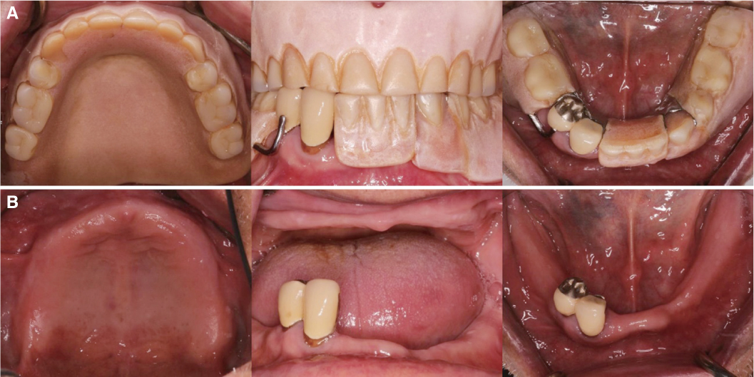
Fig. 1 Intraoral examination. (A) with existing prosthesis: Denture teeth wear and denture base resin discoloration, (B) without denture: Alveolar ridge atrophy.
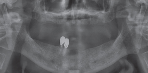
Fig. 2 Radiographic examination (Panoramic view): No condylar abnormality and intrabony lesion.
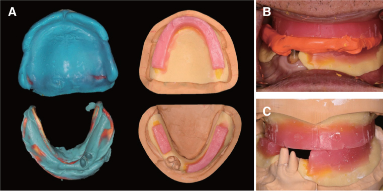
Fig. 3 (A) Final impression taking (Maxilla: Polyvinyl siloxane, Mandible: Polyether) for maxillary complete denture and mandibular surveyed crown, working cast and occlusion rim fabrication, (B) Jaw relation record, (C) Working cast mounting.
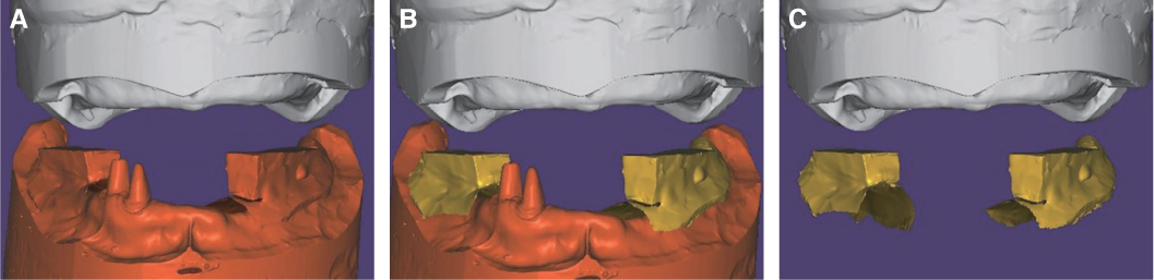
Fig. 4 Working casts scanning. (A) Maxillary and mandibular working cast, (B) Mandibular occlusal rim scan data, (C) Separation of occlusion rim scan data.
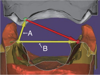
Fig. 5 Measuring the angle between interalveolar crest line (yellow line : A) and occlusal plane (yellow line : B): Occlusal rim scan data.
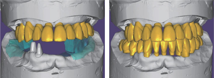
Fig. 6 Virtual teeth arrangement: Right first and second molar are set in cross bite.
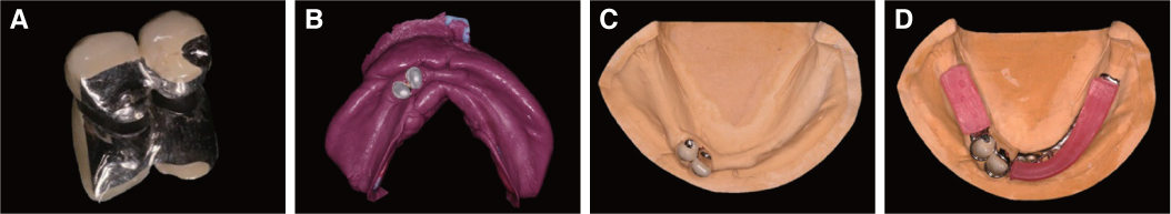
Fig. 7 7. (A) Surveyed crown fabrication, (B) Pick up impression for mandibular RPD(Polyvinyl siloxane), (C) Working cast for mandibular RPD, (D) Metal framework and occlusion rim fabrication for jaw relation record.
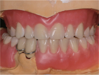
Fig. 8 Teeth arrangement and festooning: Right first and second molar are set in cross bite.
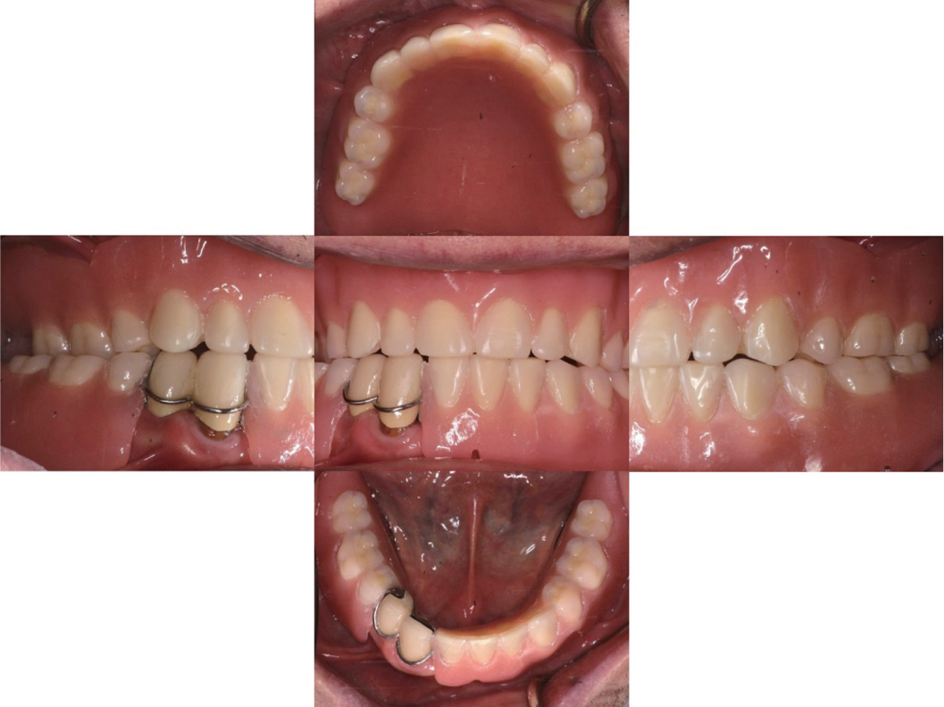
Fig. 9 Delivery of surveyed crown and removable prosthesis.
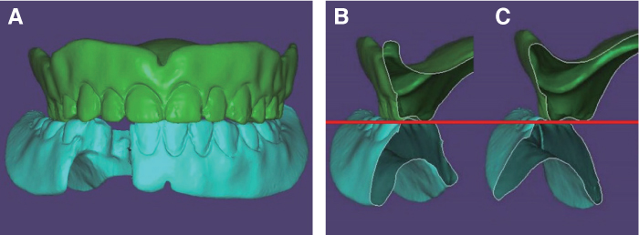
Fig. 10 (A) Scanned image of new removable prosthesis, (B) Sectional view of right second premolar: teeth are set in normal bite, (C) Sectional view of right first molar: teeth are set in cross bite. Buccal and palatal cusps show the same level.
Cited by 1 articles
-
Complete denture fabrication of a skeletal class III edentulous patient considering anterior neutral zone: a case report
Su-Hun Kim, Hyung-Jun Kim, Sang-Won Park, Hyun-Pil Lim, Chan Park, Woo-hyung Jang
J Dent Rehabil Appl Sci. 2024;40(2):91-99. doi: 10.14368/jdras.2024.40.2.91.
Reference
-
1. Atwood DA, Coy WA. Clinical, cephalometric, and densitometric study of reduction of residual ridges. J Prosthet Dent. 1971; 26:280–295.
Article2. Sanghvi SJ, Bhatt NA, Bhargava K. An evaluation of cross-bite ridge relationships. A study of articulated jaw records of 150 edentulous patients. J Prosthet Dent. 1981; 45:24–29.
Article3. Weinberg , Lawrence A. Tooth position in relation to the denture base foundation. J Prosthet Dent. 1958; 8:398–405.
Article4. Bilhan H, Geckili O, Ergin S, Erdogan O, Ates G. Evaluation of satisfaction and complications in patients with existing complete dentures. J Oral Sci. 2013; 55:29–37.
Article5. Kawahata N, Kamada Y, Ohtsuka A, Kamashita Y, Nishi Y, Hamano T, Nagaoka E. A visual method for analysing buccolingual position of artificial posterior teeth. Part 1: use of the ridge crest. J Oral Rehabil. 1998; 25:914–920.
Article6. Palla S. Occlusal considerations in complete dentures. Science and practice of occlusion. Chicago: Quintessence;1997. p. 457–467.7. Baba NZ. Materials and processes for CAD/CAM complete denture fabrication. Curr Oral Health Rep. 2016; 3:203–208.
Article8. Steinmassl O, Offermanns V, Stöckl W, Dumfahrt H, Grunert I, Steinmassl PA. In vitro analysis of the fracture resistance of CAD/CAM denture base resins. Materials (Basel). 2018; 11:E401.
Article9. Steinmassl O, Dumfahrt H, Grunert I, Steinmassl PA. CAD/CAM produces dentures with improved fit. Clin Oral Investig. 2018; 22:2829–2835.
Article10. Goodacre BJ, Goodacre CJ, Baba NZ, Kattadiyil MT. Comparison of denture tooth movement between CAD-CAM and conventional fabrication techniques. J Prosthet Dent. 2018; 119:108–115.
Article
- Full Text Links
-
- Actions
-
Cited
- CITED
-
- Close
- Share
-
- Similar articles
-
- Fixed implant rehabilitation of maxillary edentulous patient using intraoral scanning digital workflow: a case report
- Rehabilitation of maxillary partial edentulous patients using implant assisted removable partial denture
- The use of implant surveyed fixed prosthesis for removable partial denture with a few unilateral remaining teeth: A case report
- Fabrication of definitive complete-arch implant-supported fixed prosthesis in upper and lower completely edentulous patient using temporary prosthesis scan: a case report
- Complete denture fabrication of a skeletal class III edentulous patient considering anterior neutral zone: a case report
