Successful Treatment of Genital Warts with Ingenol Mebutate Monitored with Optical Coherence Tomography and Reflectance Confocal Microscopy
- Affiliations
-
- 1Department of Dermatology and Allergy, University Hospital of Munich (LMU), Munich, Germany. markus.reinholz@med.uni-muenchen.de
- 2Department of Dermatology, Okayama University Graduate School of Medicine, Dentistry and Pharmaceutical Sciences, Kawasaki Medical School, Okayama, Japan.
- KMID: 2451481
- DOI: http://doi.org/10.5021/ad.2019.31.4.434
Abstract
- Ingenol mebutate (IM) is approved for the treatment of actinic keratosis and induces cell death in precancerous lesions. The efficacy of IM in the treatment of genital warts was investigated in a therapy-refractory patient. The 74-year-old male was treated with IM gel for three consecutive days. Treatment course and efficacy were evaluated by clinical inspection and non-invasive diagnostics namely optical coherence tomography (OCT) and reflectance confocal microscopy (RCM). Within 24 to 48 hours IM induced a strong local inflammatory reaction. One week later a complete response was observed. OCT and RCM showed a strong reaction after treatment with erosions, swelling of cells, and a subepidermal dark band in representative lesions. IM has the advantage of a short treatment period in contrast to other topical treatments and shows a promising clinical outcome. Larger studies are needed to validate the data.
MeSH Terms
Figure
-
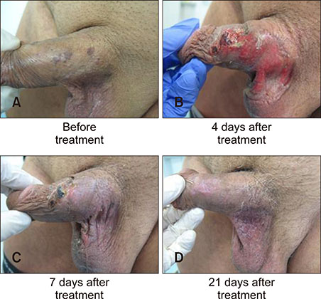
Fig. 1 Therapeutic follow-up. Patient (A) before treatment, (B) 4 days, (C) 7 days, and (D) 21 days after application of ingenol mebutate. Written informed consent to publish the photos from the patient was obtained.
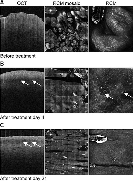
Fig. 2 Optical coherence tomography (OCT), overview reflectance confocal microscopy (RCM) mosaic, and single RCM imaging in therapeutic follow-up: (A) Patient before treatment with a verruciform lesion visible in OCT (1.8×0.6 mm, scale bar=0.5 mm) and the overview RCM mosaic (5×5 mm) at a superficial epidermal level in the single RCM image (0.5×0.5 mm) with bright epidermal cells. (B) On day 4, in OCT with a subepidermal dark band (arrows) compatible with inflammation/edema and dilated vessels in overview RCM at the level of dermoepidermal junction (DEJ), in single image RCM at the DEJ level bright particles (arrows) compatible with inflammatory cells are seen. (C) After 21 days in OCT still a subepidermal dark band (arrows) is present, in RCM vessels are still prominent (overview, at DEJ level), but epidermis shows a regular honeycomb pattern (single RCM at stratum corneum/stratum granulosum level, asterisk marks a bright artefact due to an immersion oil bubble).
Reference
-
1. Doorbar J. Molecular biology of human papillomavirus infection and cervical cancer. Clin Sci (Lond). 2006; 110:525–541.
Article2. Dietrich A, Hermans C, Heppt MV, Ruzicka T, Schauber J, Reinholz M. Human papillomavirus status, anal cytology and histopathological outcome in HIV-positive patients. J Eur Acad Dermatol Venereol. 2015; 29:2011–2018.
Article3. Rosales R, Rosales C. Immune therapy for human papillomaviruses-related cancers. World J Clin Oncol. 2014; 5:1002–1019.
Article4. Yanofsky VR, Patel RV, Goldenberg G. Genital warts: a comprehensive review. J Clin Aesthet Dermatol. 2012; 5:25–36.5. Fallen RS, Gooderham M. Ingenol mebutate: an introduction. Skin Therapy Lett. 2012; 17:1–3.6. Maier T, Braun-Falco M, Hinz T, Schmid-Wendtner MH, Ruzicka T, Berking C. Morphology of basal cell carcinoma in high definition optical coherence tomography: en-face and slice imaging mode, and comparison with histology. J Eur Acad Dermatol Venereol. 2013; 27:e97–e104.
Article7. Maier T, Braun-Falco M, Laubender RP, Ruzicka T, Berking C. Actinic keratosis in the en-face and slice imaging mode of high-definition optical coherence tomography and comparison with histology. Br J Dermatol. 2013; 168:120–128.
Article8. Reinholz M, Gauglitz GG, Giehl K, Braun-Falco M, Schwaiger H, Schauber J, et al. Non-invasive diagnosis of sweat gland dysplasia using optical coherence tomography and reflectance confocal microscopy in a family with anhidrotic ectodermal dysplasia (Christ-Siemens-Touraine syndrome). J Eur Acad Dermatol Venereol. 2016; 30:677–682.
Article9. Maier T, Cekovic D, Ruzicka T, Sattler EC, Berking C. Treatment monitoring of topical ingenol mebutate in actinic keratoses with the combination of optical coherence tomography and reflectance confocal microscopy: a case series. Br J Dermatol. 2015; 172:816–818.
Article10. von Braunmühl T, Welzel J. [Noninvasive diagnostic imaging in dermatology]. Hautarzt. 2015; 66:492. German.11. von Braunmühl T. [Optical coherence tomography]. Hautarzt. 2015; 66:499–503. German.12. Anderson L, Schmieder GJ, Werschler WP, Tschen EH, Ling MR, Stough DB, et al. Randomized, double-blind, double-dummy, vehicle-controlled study of ingenol mebutate gel 0.025% and 0.05% for actinic keratosis. J Am Acad Dermatol. 2009; 60:934–943.
Article13. von Krogh G, Szpak E, Andersson M, Bergelin I. Self-treatment using 0.25%–0.50% podophyllotoxin-ethanol solutions against penile condylomata acuminata: a placebo-controlled comparative study. Genitourin Med. 1994; 70:105–109.
Article14. Schöfer H, Van Ophoven A, Henke U, Lenz T, Eul A. Randomized, comparative trial on the sustained efficacy of topical imiquimod 5% cream versus conventional ablative methods in external anogenital warts. Eur J Dermatol. 2006; 16:642–648.15. Gross G, Meyer KG, Pres H, Thielert C, Tawfik H, Mescheder A. A randomized, double-blind, four-arm parallel-group, placebo-controlled Phase II/III study to investigate the clinical efficacy of two galenic formulations of Polyphenon E in the treatment of external genital warts. J Eur Acad Dermatol Venereol. 2007; 21:1404–1412.
Article16. Khawaja HT. Treatment of condyloma acuminatum. Lancet. 1986; 1:208–209.
Article17. Khawaja HT. Podophyllin versus scissor excision in the treatment of perianal condylomata acuminata: a prospective study. Br J Surg. 1989; 76:1067–1068.
Article18. Braun SA, Jansen TM, Homey B, Gerber PA. [Successful therapy of condylomata acuminata with ingenol mebutate]. Hautarzt. 2015; 66:223–225. German.
Article19. Rosen RH, Gupta AK, Tyring SK. Dual mechanism of action of ingenol mebutate gel for topical treatment of actinic keratoses: rapid lesion necrosis followed by lesion-specific immune response. J Am Acad Dermatol. 2012; 66:486–493.
Article20. Ogbourne SM, Suhrbier A, Jones B, Cozzi SJ, Boyle GM, Morris M, et al. Antitumor activity of 3-ingenyl angelate: plasma membrane and mitochondrial disruption and necrotic cell death. Cancer Res. 2004; 64:2833–2839.
- Full Text Links
-
- Actions
-
Cited
- CITED
-
- Close
- Share
-
- Similar articles
-
- A Case of Molluscum Contagiosum Treated by Ingenol Mebutate (Picato®)
- Successful Treatment of Bowenoid Papulosis with Fractional CO2 Laser and Ingenol Mebutate Gel
- The Findings of Reflectance Confocal Microscopy in the Diagnosis of Green Nail Syndrome
- Inflammatory Vitiligo Confirmed Diagnosis and Treatment Outcome Using a Reflectance Confocal Microscopy
- Clinical Experience of Ingenol Mebutate Therapy in Extramammary Paget Disease Accompanying Condyloma Acuminatum
