In vivo magnetic resonance imaging morphometry of the patella bone in South Indian population
- Affiliations
-
- 1Department of Anatomy, Kasturba Medical College, Manipal University, Mangalore, India. vasudha.vs@manipal.edu
- KMID: 2451234
- DOI: http://doi.org/10.5115/acb.2017.50.2.99
Abstract
- Racial differences exist in the dimensions of structures and the commercially available prostheses are designed based on the Caucasians. In this context, the goal of the present investigation was to determine the gender wise measurements of patella bone in South Indians. The present study included axial magnetic resonance images of the knee joint from 140 South Indian adults (70 males, 70 females; aged between 20-70 years). The angle, width, thickness, lateral facet width, facet thickness, ratio of the lateral facet, the relative thickness and ratio of facet thickness were measured in the patella by using the digital ruler. The statistical analysis was performed by using the SPSS software. The dimensions exhibited statistically highly significant sexual dimorphism (P≤0.001). The mean value was higher in males than females except for the ratio of patellar lateral facet and patellar facet thickness ratio. It was observed that the males exhibit more variability than females in all the measurements of patella except patellar thickness, patellar facet thickness, patellar relative thickness, and patellar facet thickness ratio. The present study of the in vivo morphometry of patella bone from the South Indians can provide a population and gender specific database for the morphometric measurements of the patella. We believe that the data of the present study will be useful to the orthopaedician during the procedures like arthroplasty of the total knee, patellofemoral arthroplasty, resurfacing of patella, and designing the prosthetic implant.
MeSH Terms
Figure
-
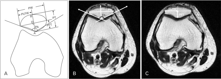
Fig. 1 The schematic representation (A) (A, point of the patellar central ridge; B, patellar anterior point; O, patellar central point) and the magnetic resonance imaging (MRI) right knee T2 axial section (B) illustrating the landmarks for measuring the patella angle (PA), the patella width (PW), and the patella thickness (PT). The MRI right knee T2 axial section (C) illustrates the landmarks for measuring patella lateral facet width (PLFW) and the patella facet thickness (PFT).
Cited by 2 articles
-
Morphometric analysis of vastus medialis oblique muscle and its influence on anterior knee pain
Marwa M El Sawy, Dalia M E EL Mikkawy, Sayed M El-Sayed, Ahmed M. Desouky
Anat Cell Biol. 2021;54(1):1-9. doi: 10.5115/acb.20.258.Assessment of thickness of in vivo autograft tendons around the knee and its correlation with anthropometric data, thickness of patella and anterior cruciate ligament tibial foot print diameter
Balgovind S Raja, Kshitij Gupta, Abdusamad V, Sukhmin Singh, Subhajit Maji
Anat Cell Biol. 2021;54(1):18-24. doi: 10.5115/acb.20.176.
Reference
-
1. Faraj AA, Nevelos AB. Ethnic factors in Perthes disease: a retrospective study among white and Asian population living in the same environment. Acta Orthop Belg. 2000; 66:255–258. PMID: 11033915.2. Ho WP, Cheng CK, Liau JJ. Morphometrical measurements of resected surface of femurs in Chinese knees: correlation to the sizing of current femoral implants. Knee. 2006; 13:12–14. PMID: 16122927.3. Kwak DS, Han S, Han CW, Han SH. Resected femoral anthropometry for design of the femoral component of the total knee prosthesis in a Korean population. Anat Cell Biol. 2010; 43:252–259. PMID: 21212865.4. Lim HC, Bae JH, Yoon JY, Kim SJ, Kim JG, Lee JM. Gender differences of the morphology of the distal femur and proximal tibia in a Korean population. Knee. 2013; 20:26–30. PMID: 22721912.5. Vaidya SV, Ranawat CS, Aroojis A, Laud NS. Anthropometric measurements to design total knee prostheses for the Indian population. J Arthroplasty. 2000; 15:79–85. PMID: 10654467.6. Yang B, Tan H, Yang L, Dai G, Guo B. Correlating anatomy and congruence of the patellofemoral joint with cartilage lesions. Orthopedics. 2009; 32:20. PMID: 19226043.7. Shang P, Zhang L, Hou Z, Bai X, Ye X, Xu Z, Huang X. Morphometric measurement of the patella on 3D model reconstructed from CT scan images for the southern Chinese population. Chin Med J (Engl). 2014; 127:96–101. PMID: 24384431.8. Introna F Jr, Di Vella G, Campobasso CP. Sex determination by discriminant analysis of patella measurements. Forensic Sci Int. 1998; 95:39–45. PMID: 9718670.9. Baldwin JL, House CK. Anatomic dimensions of the patella measured during total knee arthroplasty. J Arthroplasty. 2005; 20:250–257. PMID: 15902866.10. Hitt K, Shurman JR 2nd, Greene K, McCarthy J, Moskal J, Hoeman T, Mont MA. Anthropometric measurements of the human knee: correlation to the sizing of current knee arthroplasty systems. J Bone Joint Surg Am. 2003; 85-A(Suppl 4):115–122. PMID: 12533581.11. Kim TK, Chung BJ, Kang YG, Chang CB, Seong SC. Clinical implications of anthropometric patellar dimensions for TKA in Asians. Clin Orthop Relat Res. 2009; 467:1007–1014. PMID: 18855087.12. Borotikar BS, Sipprell WH 3rd, Wible EE, Sheehan FT. A methodology to accurately quantify patellofemoral cartilage contact kinematics by combining 3D image shape registration and cine-PC MRI velocity data. J Biomech. 2012; 45:1117–1122. PMID: 22284428.13. Iranpour F, Merican AM, Amis AA, Cobb JP. The width:thickness ratio of the patella: an aid in knee arthroplasty. Clin Orthop Relat Res. 2008; 466:1198–1203. PMID: 18330664.14. Yoo JH, Yi SR, Kim JH. The geometry of patella and patellar tendon measured on knee MRI. Surg Radiol Anat. 2007; 29:623–628. PMID: 17898923.15. McHale PL, McGarry MH, LaRoque ES, Schulz MM, Lee TQ. Gender differences in the patellofemoral joint: effects of lateral release. In : Transactions of the 48th Annual Meeting of the Orthopaedic Research Society; 2002 Feb 10-13; Dallas, Taxas. Poster No. 898.16. Hsu HC, Luo ZP, Rand JA, An KN. Influence of patellar thickness on patellar tracking and patellofemoral contact characteristics after total knee arthroplasty. J Arthroplasty. 1996; 11:69–80. PMID: 8676121.17. Bogen H. Uber familiiire Luxation und Rleinheit der Pat. Z Othop Chir. 1906; 16:359–419.18. Akhlaghi M, Sheikhazadi A, Naghsh A, Dorvashi G. Identification of sex in Iranian population using patella dimensions. J Forensic Leg Med. 2010; 17:150–155. PMID: 20211456.19. Bidmos MA, Steinberg N, Kuykendall KL. Patella measurements of South African whites as sex assessors. Homo. 2005; 56:69–74. PMID: 15901119.20. Dayal MR, Bidmos MA. Discriminating sex in South African blacks using patella dimensions. J Forensic Sci. 2005; 50:1294–1297. PMID: 16382821.21. Kemkes-Grottenthaler A. Sex determination by discriminant analysis: an evaluation of the reliability of patella measurements. Forensic Sci Int. 2005; 147:129–133. PMID: 15567616.
- Full Text Links
-
- Actions
-
Cited
- CITED
-
- Close
- Share
-
- Similar articles
-
- In vivo magnetic resonance imaging morphometry of the patella bone in South Indian population
- Morphometric study of tensor of vastus intermedius in South Indian population
- Preoperative Templating of Bone-Patellar Tendon-Bone Graft for Anterior Cruciate Ligament Reconstruction: A Morphometry-Based Graft Harvest Method
- Sleeve Fracture of the Superior Pole of Patella in an Adolescent
- Idiopathic Osteonecrosis of the Patella: A Case Report
