Morphometric study of tensor of vastus intermedius in South Indian population
- Affiliations
-
- 1Department of Anatomy, Jawaharlal Institute of Postgraduate Medical Education and Research (JIPMER), Pondicherry, India. dr_raveendra@rediffmail.com
- KMID: 2390420
- DOI: http://doi.org/10.5115/acb.2017.50.1.7
Abstract
- Tensor of vastus intermedius is a newly discovered muscle located between vastus lateralis and vastus intermedius. The purpose of this study was to investigate the detailed morphology of tensor of vastus intermedius, specifically to provide data pertaining to the attachments, innervations, variation in the types and its morphometry in South Indian population. The tensor of vastus intermedius was studied in thirty six cadaveric lower limbs using macrodissection techniques. The origin of the muscle was from upper part of intertrochanteric line and anterior part of greater trochanter of femur inserted to medial aspect of upper border of patella. The muscle was classified into four types based on the origin and also the aponeurosis course with independent type (type 1) being common. The mean and standard deviation of the length of tensor of vastus intermedius and aponeurosis were 145.40±37.55 mm and 193.55±42.32 mm, respectively. The results of the study suggest that tensor of vastus intermedius is variable and the information provided regarding the attachments, types and quantitative data will contribute to the existing knowledge of the muscle.
Figure
-
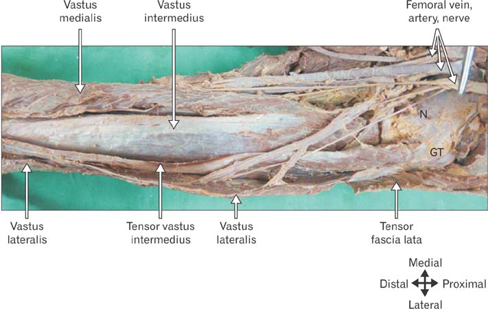
Fig. 1 Anterior view of left thigh. Tensor of vastus intermedius origins from the upper part of intertrochanteric line and anterior part of greater trochanter. It lies between vastus lateralis and vastus intermedius throughout its entire course. The anterior border of tensor of vastus intermedius in the proximal part becomes medial border distally. Distally the lateral border of tensor of vastus intermedius fuses with vastus lateralis. The muscle is supplied by branches from femoral vessels and nerves. N, neck of femur; GT, greater trochanter of femur.
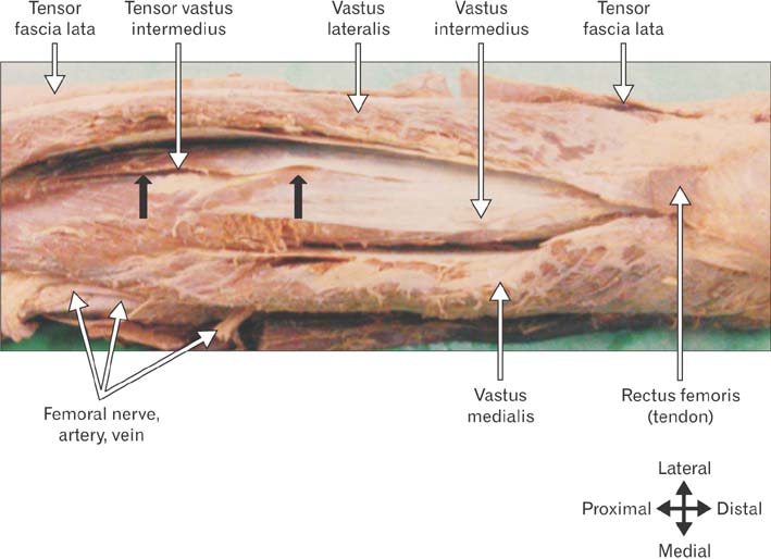
Fig. 2 Anterior view of left thigh. Rectus femoris is incised and reflected proximally, so not seen. Tensor of vastus intermedius arises along with vastus intermedius. The posterior border of aponeurosis is fused with vastus intermedius.
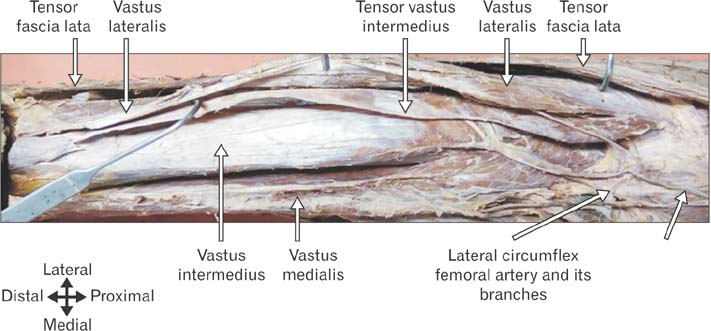
Fig. 3 Anterior view of right thigh. Rectus femoris is incised and reflected proximally, so not seen. The tensor of vastus intermedius arises along with vastus lateralis. The aponeurosis is separable from vastus intermedius and not from vastus lateralis.
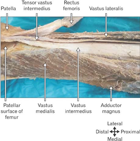
Fig. 4 Anterior view of right thigh. Rectus femoris is reflected laterally. The attachment of vastus medialis to patella is incised. The tensor of vastus intermedius aponeurosis inserted into the upper border of patella in the intermediate layer between rectus femoris above and vastus intermedius below.
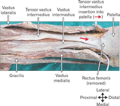
Fig. 5 Anterior view of left thigh. Rectus femoris is incised near its insertion into patella base. The tensor of vastus intermedius aponeurosis runs medially and inserts into the base of patella.
Reference
-
1. Grob K, Ackland T, Kuster MS, Manestar M, Filgueira L. A newly discovered muscle: the tensor of the vastus intermedius. Clin Anat. 2016; 29:256–263.2. Willan PL, Mahon M, Golland JA. Morphological variations of the human vastus lateralis muscle. J Anat. 1990; 168:235–239.3. Lin F, Wang G, Koh JL, Hendrix RW, Zhang LQ. In vivo and noninvasive three-dimensional patellar tracking induced by individual heads of quadriceps. Med Sci Sports Exerc. 2004; 36:93–101.4. Miao P, Xu Y, Pan C, Liu H, Wang C. Vastus medialis oblique and vastus lateralis activity during a double-leg semisquat with or without hip adduction in patients with patellofemoral pain syndrome. BMC Musculoskelet Disord. 2015; 16:289.5. Lefebvre R, Leroux A, Poumarat G, Galtier B, Guillot M, Van-neuville G, Boucher JP. Vastus medialis: anatomical and functional considerations and implications based upon human and cadaveric studies. J Manipulative Physiol Ther. 2006; 29:139–144.6. Goh JC, Lee PY, Bose K. A cadaver study of the function of the oblique part of vastus medialis. J Bone Joint Surg Br. 1995; 77:225–231.7. Grob K, Fretz C, Kuster MS, Gilbey H, Ackland T. Knee pain associated with rupture of tensor vastus intermedius, a newly discovered muscle: a case report. J Clin Case Rep. 2016; 6:828.8. Rajasekaran S, Hall MM. Sonographic appearance of the tensor of the vastus intermedius. PM R. 2016; 8:1020–1023.
- Full Text Links
-
- Actions
-
Cited
- CITED
-
- Close
- Share
-
- Similar articles
-
- Muscle Thickness and Echo Intensity of the Abdominal and Lower Extremity Muscles in Stroke Survivors
- A Computed Tomography-Based Assessment of the Anatomical Parameters Concerning S2-Alar Iliac Screw Insertion Using “Safe Trajectory Method” in Indian Population
- Exploring the atlantic part of the vertebral artery in the South Indian population and its implications in spine surgery
- Computed Tomography-Based Occipital Condyle Morphometry in an Indian Population to Assess the Feasibility of Condylar Screws for Occipitocervical Fusion
- Morphometric Study of C1 Pedicle and Feasibility Evaluation of C1 Pedicle Screw Placement with a Novel Clinically Relevant Radiological Classification in an Indian Population
