Anat Cell Biol.
2019 Jun;52(2):214-216. 10.5115/acb.2019.52.2.214.
Drainage of the basal vein of Rosenthal into the confluence of sinuses
- Affiliations
-
- 1Department of Anatomical Sciences, St. George's University, St. George's, Grenada, West Indies.
- 2Seattle Science Foundation, Seattle, WA, USA. joei@seattlesciencefoundation.org
- 3Division of Neuroradiogy, University of Alabama, Birmingham, AL, USA.
- KMID: 2451228
- DOI: http://doi.org/10.5115/acb.2019.52.2.214
Abstract
- An adult female was found to have a variation of the left basal vein of Rosenthal after presenting with complaints of headache. The vein, in this case, drained directly into the confluence of sinuses instead of the great vein of Galen. Variation of the basal vein is likely due to the embryonic development of the deep cerebral venous system as primitive structures either differentiate further or regress with age. Such changes may result in the uncommon presentation seen in this case. To our knowledge, this is the first case reported of the basal vein draining into the confluence of sinuses.
Figure
-
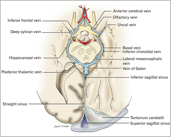
Fig. 1 Schematic drawing of the normal course of the basal veins of Rosenthal.
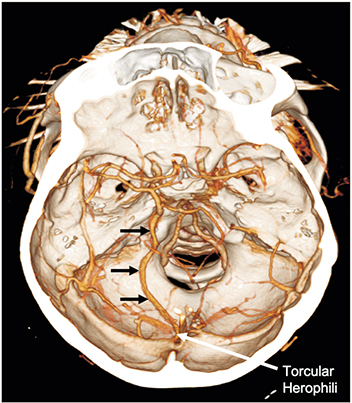
Fig. 2 Computed tomography angiography of the patient presented herein noting the left basal vein of Rosenthal (arrows) draining posteriorly into the confluence of sinuses.
Reference
-
1. Tubbs RS, Loukas M, Louis RG Jr, Shoja MM, Askew CS, Phantana-Angkool A, Salter EG, Oakes WJ. Surgical anatomy and landmarks for the basal vein of rosenthal. J Neurosurg. 2007; 106:900–902.
Article2. Padget DH. The development of the cranial venous system in man: from the viewpoint of comparative anatomy. Baltimore, MD: Carnegie Institution of Washington;1957.3. Suzuki Y, Ikeda H, Shimadu M, Ikeda Y, Matsumoto K. Variations of the basal vein: identification using three-dimensional CT angiography. AJNR Am J Neuroradiol. 2001; 22:670–676.4. Chung JI, Weon YC. Anatomic variations of the deep cerebral veins, tributaries of basal vein of rosenthal: embryologic aspects of the regressed embryonic tentorial sinus. Interv Neuroradiol. 2005; 11:123–130.
Article5. Watanabe A, Hirano K, Kamada M, Imamura K, Ishii N, Sekihara Y, Suzuki Y, Ishii R. Perimesencephalic nonaneurysmal subarachnoid haemorrhage and variations in the veins. Neuroradiology. 2002; 44:319–325.
Article6. Shoja MM, Ramdhan R, Jensen CJ, Chern JJ, Oakes WJ, Tubbs RS. Embryology of the craniocervical junction and posterior cranial fossa, part II: embryogenesis of the hindbrain. Clin Anat. 2018; 31:488–500.
Article7. George Zaki Ghali M. Galenic pial arteriovenous fistulas: angioarchitecture, clinical presentation, and therapeutic considerations. Clin Anat. 2018; 31:259–268.
Article
- Full Text Links
-
- Actions
-
Cited
- CITED
-
- Close
- Share
-
- Similar articles
-
- Direct drainage of the basal vein of Rosenthal into the superior petrosal sinus: a literature review
- A Unique Type of Dural Arteriovenous Fistula at Confluence of Sinuses Treated with Endovascular Embolization: A Case Report
- Significance of Cerebral Venography in Surgery of Petroclival Meningiomas
- The Experience of Ligation of Transverse or Sigmoid Sinus in Surgery of Large Petroclival Meningiomas
- Measurements of basal vein of Rosenthal in normal adult
