A Case of Familial Spondyloenchondrodysplasia with Immune Dysregulation Masquerading as Moyamoya Syndrome
- Affiliations
-
- 1Department of Pediatrics, Department of Genome Medicine and Science, Gil Medical Center, Gachon University College of Medicine, Incheon, Korea.
- 2Department of Pediatrics, Pediatric Clinical Neuroscience Center, Seoul National University Children's Hospital, Seoul National University College of Medicine, Seoul, Korea. chaeped1@snu.ac.kr
- 3Division of Pediatric Neurosurgery, Seoul National University Children's Hospital, Seoul National University College of Medicine, Seoul, Korea.
- 4Department of Radiology, Woorisoa Children's Hospital, Seoul, Korea.
- 5Department of Pediatrics, Seoul National University Children's Hospital, Seoul National University College of Medicine, Seoul, Korea.
- 6Cell Logistics Research Center and School of Life Sciences, Gwangju Institute of Science and Technology, Gwangju, Korea.
- 7Division of Pediatric Orthopaedics, Seoul National University Children's Hospital, Seoul National University College of Medicine, Seoul, Korea.
- KMID: 2451130
- DOI: http://doi.org/10.3988/jcn.2019.15.3.407
Abstract
- No abstract available.
MeSH Terms
Figure
-
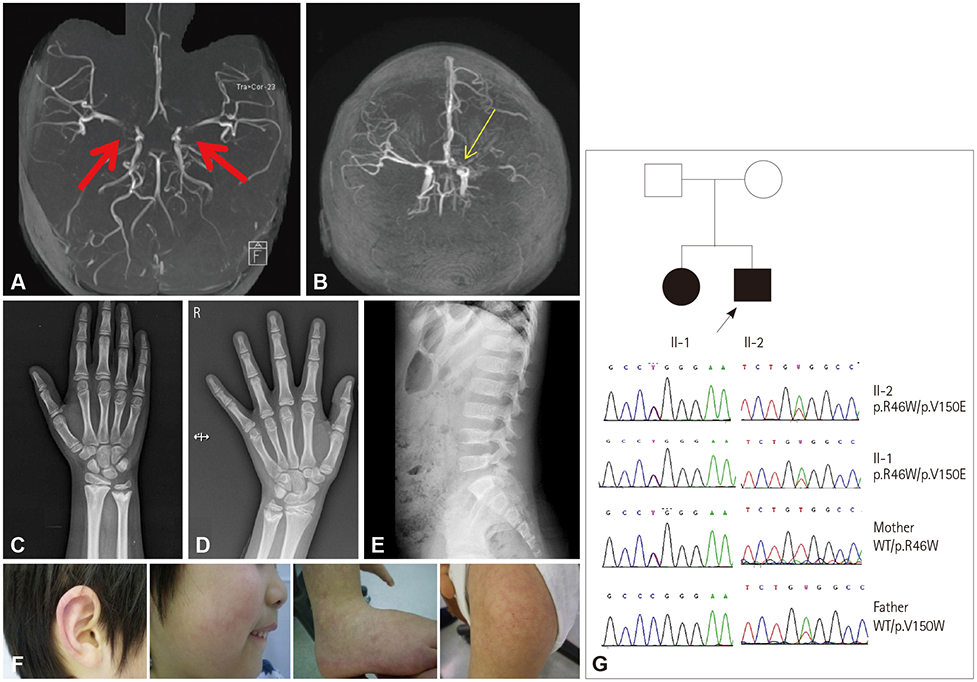
Fig. 1 Radiology, photograph and Sanger sequencing. Magnetic resonance angiography images of the brain show bilateral stenosis/occlusion (red arrows) of the proximal middle and anterior cerebral arteries in the proband (II-2) (A) and complete occlusion (yellow arrow) of the left middle and anterior cerebral arteries and focal narrowing of the right proximal middle cerebral artery in his sister (II-1) (B). Radiographic photographs of the proband show bilateral metaphyseal irregularities with cupped and enchondromata-like streaks in the radius and ulna (C and D), and generalized platyspondyly of the vertebral bodies with a rectangular shape (E). Skin lesions of the proband were suggestive of Raynaud's phenomenon (F). Pedigrees and Sanger sequencing revealed compound heterozygous variants of ACP5 in both affected siblings: c.136C>T (p.R46W) and c.449T>A (p.V150E). The black arrow indicates the proband (II-2) (G). WT: wild type.
Reference
-
1. Schorr S, Legum C, Ochshorn M. Spondyloenchondrodysplasia. Enchondromatomosis with severe platyspondyly in two brothers. Radiology. 1976; 118:133–139.2. Frydman M, Bar-Ziv J, Preminger-Shapiro R, Brezner A, Brand N, Ben-Ami T, et al. Possible heterogeneity in spondyloenchondrodysplasia: quadriparesis, basal ganglia calcifications, and chondrocyte inclusions. Am J Med Genet. 1990; 36:279–284.
Article3. Briggs TA, Rice GI, Adib N, Ades L, Barete S, Baskar K, et al. Spondyloenchondrodysplasia due to mutations in ACP5: a comprehensive survey. J Clin Immunol. 2016; 36:220–234.
Article4. Renella R, Schaefer E, LeMerrer M, Alanay Y, Kandemir N, Eich G, et al. Spondyloenchondrodysplasia with spasticity, cerebral calcifications, and immune dysregulation: clinical and radiographic delineation of a pleiotropic disorder. Am J Med Genet A. 2006; 140:541–550.
Article5. Bae JS, Kim NK, Lee C, Kim SC, Lee HR, Song HR, et al. Comprehensive genetic exploration of skeletal dysplasia using targeted exome sequencing. Genet Med. 2016; 18:563–569.
Article6. Navarro V, Scott C, Briggs TA, Barete S, Frances C, Lebon P, et al. Two further cases of spondyloenchondrodysplasia (SPENCD) with immune dysregulation. Am J Med Genet A. 2008; 146A:2810–2815.
Article7. Wang R, Xu Y, Lv R, Chen J. Systemic lupus erythematosus associated with Moyamoya syndrome: a case report and literature review. Lupus. 2013; 22:629–633.
Article8. Jeong HC, Kim YJ, Yoon W, Joo SP, Lee SS, Park YW. Moyamoya syndrome associated with systemic lupus erythematosus. Lupus. 2008; 17:679–682.
Article
- Full Text Links
-
- Actions
-
Cited
- CITED
-
- Close
- Share
-
- Similar articles
-
- Spondyloenchondrodysplasia with immune dysregulation: an under-the-radar cause of spasticity
- A Case of Familial Moyamoya Disease: A Case in Brother and Sister
- Familial Occurrence of Moyamoya Disease in a Father and a Son
- Familial Occurrence of Moyamoya Disease: Case Report
- Systemic Lupus Erythematosus Associated with Familial Moyamoya Disease
