Imaging Sci Dent.
2019 Jun;49(2):153-158. 10.5624/isd.2019.49.2.153.
Morphological variation of the velum in children and adults using magnetic resonance imaging
- Affiliations
-
- 1Department of Communication Sciences and Disorders, East Carolina University, Greenville, NC, USA. perryja@ecu.edu
- KMID: 2450181
- DOI: http://doi.org/10.5624/isd.2019.49.2.153
Abstract
- PURPOSE
The purpose of this study was to investigate variations in velar shape according to age, sex, and race using magnetic resonance imaging (MRI).
MATERIALS AND METHODS
The study sample consisted of 170 participants (85 children, 85 adults) between 4 and 34 years of age. Velar morphology was visually classified using midsagittal MRI scans for each participant by 2 independent raters. Inter- and intra-rater reliability was assessed. Statistical analyses were performed to identify correlations of velar shape with sex, age, and race.
RESULTS
The most frequent velar shape was "buttf" for both adults (41%) and children (58%) in this study. The least common shapes for adults were "leaf" and "S.f" The children did not exhibit any "leaff" or "straightf" velar shapes. A statistically significant difference was noted for age with respect to velar shape (P=0.014). Sex and race were found to have no significant impact on velar shape in this study.
CONCLUSION
When using MRI to evaluate velar morphology, the "buttf" shape was most common in both children and adults. Velar shape varied significantly with age, while race and sex did not have a significant impact.
Keyword
MeSH Terms
Figure
-
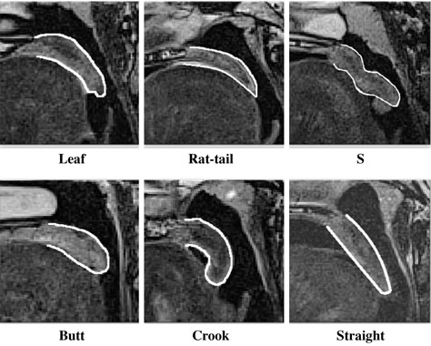
Fig. 1 Midsagittal magnetic resonance images of adults show all 6 morphological categories of velar shape. The velum is outlined in white to emphasize its shape and to differentiate it from the surrounding structures.
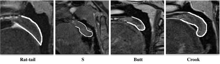
Fig. 2 Midsagittal magnetic resonance images of children show 4 morphological categories of velar shape. The velum is outlined in white to emphasize its shape and to differentiate it from the surrounding structures. The “leaf” and “straight” shapes are not shown because there were no instances of these types within the sample of children analyzed in this study.
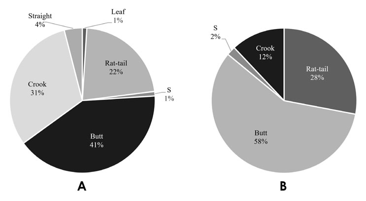
Fig. 3 Pie charts depicting the distribution of each velar shape category in adults (A) and children (B).
Reference
-
1. Deepa V, David CM, Ramnarayan BK. Morphological varieties of soft palate in normal individuals, cleft palate patients and obstructive sleep apnea patients with reference to Indian population: a preliminary digital cephalometric study. World J Dent. 2013; 4:241–249.
Article2. Deshmukh RA, Bagewadi AS. Morphometric evaluation and comparison of soft palate in individuals with and without oral submucous fibrosis: a digital cephalometric study. SRM J Res Dent Sci. 2015; 6:220–224.
Article3. Raja Lakshmi C, Ayesha Thabusum D, Bhavana SM. An innovative approach to evaluate the morphological patterns of soft palate in oral submucous fibrosis patients: a digital cephalometric study. Int J Chronic Dis. 2016; 2016:5428581.
Article4. Mohan RS, Verma S, Singh U, Agarwal N. Morphometric evaluation of soft palate in oral submucous fibrosis - a digital cephalometric analysis. West Afr J Radiol. 2014; 21:7–11.5. Pépin JL, Veale D, Ferretti GR, Mayer P, Lévy PA. Obstructive in awake patients - cephalometric and CT findings. Radiology. 1999; 210:163–170.6. You M, Li X, Wang H, Zhang J, Wu H, Liu Y, et al. Morphological variety of the soft palate in normal individuals: a digital cephalometric study. Dentomaxillofac Radiol. 2008; 37:344–349.
Article7. Guttal KS, Breh R, Bhat R, Burde KN, Naikmasur VG. Diverse morphologies of soft palate in normal individuals: a cephalometric perspective. J Indian Acad Oral Med Radiol. 2012; 24:15–19.8. Kumar DK, Gopal DK. Morphological variants of soft palate in normal individuals: a digital cephalometric study. J Clin Diagn Res. 2011; 5:1310–1313.9. Nagaraj T, Goswami RD, James L, Sreelakshmi N, Veerabasavaiah BT, Shruthi R. A radiographic assessment of morphologies of soft palate: a retrospective study. J Med Radiol Pathol Surg. 2016; 3:1–4.
Article10. Niu YM, Wang H, Zheng Q, He X, Zhang J, Li XM, et al. Morphology of the soft palate in normal humans with digital cephalometry. Hua Xi Kou Qiang Yi Xue Za Zhi. 2006; 24:321–327.11. Praveen BN, Amrutesh S, Pal S, Shubhasini AR, Vaseemuddin S. Various shapes of soft palate: a lateral cephalometric study. World J Dent. 2011; 2:207–210.
Article12. Santosh VK, Singh P, Pagare SS. Normative soft palate dimensions and morphology in a subset of Indian population: a digital cephalometric study. Indian J Oral Health Res. 2015; 1:48–51.
Article13. Shankar VN, Hegde K, Ashwini NS, Praveena V, Ravi Prakash SM. Morphometric evaluation of soft palate in oral submucous fibrosis - a digital cephalometric study. J Craniomaxillofac Surg. 2014; 42:48–52.14. Verma P, Verma KG, Kumaraswam KL, Basavaraju S, Sachdeva SK, Juneja S. Correlation of morphological variants of the soft palate and Need's ratio in normal individuals: a digital cephalometric study. Imaging Sci Dent. 2014; 44:193–198.
Article15. Khaitan T, Pachigolla R, Uday G, Balmuri PK, Chennoju SK, Pattipati S. Digital cephalometric analysis illustrating morphological variation of the soft palate. J Indian Acad Oral Med Radiol. 2015; 27:532–538.
Article16. Agrawal P, Gupta A, Phulambrikar T, Singh SK, Sharma BK, Rodricks D. A focus on variation in morphology of soft palate using cone-beam computed tomography with assessment of Need's ratio in central Madhya Pradesh population. J Clin Diagn Res. 2016; 10:ZC68–ZC71.
Article17. More CB, Saha N, Vijayvargiya R. Morphological study of soft palate by using computed tomography - a prospective study. J Clin Diagn Res. 2015; 9:ZC71–ZC74.18. Perry JL, Kuehn DP, Sutton BP, Gamage JK, Fang X. Anthropometric analysis of the velopharynx and related craniometric dimensions in three adult populations using MRI. Cleft Palate Craniofac J. 2016; 53:e1–e13.
Article19. U.S. Food and Drug Administration [Internet]. Silver Spring, MD: The Administration;cited 2019 Jan 31. Benefits and risks. Available from: https://www.fda.gov/Radiation-Emitting-Products/RadiationEmittingProductsandProcedures/MedicalImaging/MRI/ucm482765.htm.20. Bae Y, Kuehn DP, Sutton BP, Conway CA, Perry JL. Three-dimensional magnetic resonance imaging of velopharyngeal structures. J Speech Lang Hear Res. 2011; 54:1538–1545.
Article21. Perry JL, Sutton BP, Kuehn DP, Gamage JK. Using MRI for assessing velopharyngeal structures and function. Cleft Palate Craniofac J. 2014; 51:476–485.
Article22. Mason KN, Perry JL, Riski JE, Fang X. Age-related changes between the level of velopharyngeal closure and the cervical spine. J Craniofac Surg. 2016; 27:498–503.
Article23. Perry JL, Kollara L, Kuehn DP, Sutton BP, Fang X. Examining age, sex, and race characteristics of velopharyngeal structures in 4- to 9-year-old children using magnetic resonance imaging. Cleft Palate Craniofac J. 2018; 55:21–34.
Article24. Senaratna CV, Perret JL, Lodge CJ, Lowe AJ, Campbell BE, Matheson MC, et al. Prevalence of obstructive sleep apnea in the general population: a systematic review. Sleep Med Rev. 2017; 34:70–81.
Article25. Bixler EO, Vgontzas AN, Lin HM, Liao D, Calhoun S, Vela-Bueno A, et al. Sleep disordered breathing in children in a general population sample: prevalence and risk factors. Sleep. 2009; 32:731–736.
Article26. Perry JL. Variations in velopharyngeal structures between upright and supine positions using upright magnetic resonance imaging. Cleft Palate Craniofac J. 2011; 48:123–133.
Article27. Kollara L, Perry JL. Effects of gravity on the velopharyngeal structures in children using upright magnetic resonance imaging. Cleft Palate Craniofac J. 2014; 51:669–676.
Article
- Full Text Links
-
- Actions
-
Cited
- CITED
-
- Close
- Share
-
- Similar articles
-
- Advanced Magnetic Resonance Imaging for Pediatric Brain Tumors: Current Imaging Techniques and Interpretation Algorithms
- Ultrafast Magnetic Resonance Imaging: Echo Planar Imaging and Spiral Scan Imaging
- Morphological Analysis of the Cervical Spinal Cord, Dural Tube, and Spinal Canal by Magnetic Resonance Imaging in Normal Korean Adults
- MR Imaging and Ultrasonographic Findings of Tensor Fasciae Suralis Muscle: A Case Report
- Dorsoscapularis triangularis: embryological and phylogenetic characterization of a rare variation of trapezius
