Corneal Ulcer Caused by Corynebacterium macginleyi
- Affiliations
-
- 1Department of Ophthalmology, Chonbuk National University Medical School, Jeonju, Korea. you2ic@daum.net
- 2Research Institute of Clinical Medicine, Chonbuk National University, Jeonju, Korea.
- 3Biomedical Research Institute, Chonbuk National University Hospital, Jeonju, Korea.
- KMID: 2449712
- DOI: http://doi.org/10.3341/jkos.2019.60.6.582
Abstract
- PURPOSE
To report a case of a Corynebacterium macginleyi-infected corneal ulcer of a patient who had been treated for conjunctivitis for more than 3 months.
CASE SUMMARY
A 72-year-old female was transferred from a private ophthalmic clinic for evaluation of herpetic keratitis with progressive corneal edema and infiltration in the left eye. She had a history of conjunctival hyperemia and eyeball pain in her left eye 3 months prior to her visit. She was treated with levofloxacin eye drops and acyclovir ointment (Herpesid®, Samil, Co., Ltd. Seoul, Korea). On slit lamp examination, 5.4 × 4.0 mm corneal epithelial defects and stromal infiltrations were observed in the upper to central cornea, and endothelial keratic precipitates were found. Gram positive bacteria were detected on Gram staining and Corynebacterium macginleyi was identified on bacterial cultures from the conjunctiva and cornea. She was treated with topical vancomycin eye drops. After 3 months of treatment, the corneal ulcer was completely resolved, leaving mild superficial opacity on the cornea.
CONCLUSIONS
While Corynebacterium macginleyi, normal flora of the conjunctiva, is considered a major causative agent for conjunctivitis and blepharitis, Corynebacterium macginleyi should also be considered a possible cause of slowly progressive keratitis in patients with chronic conjunctivitis.
MeSH Terms
Figure
-

Figure 1 Slit-lamp photograph of the left eye at the first visit. (A) Deep corneal stromal infiltration and diffuse corneal edema were seen. (B) Corneal epithelial defects in the fluorescein staining were observed.

Figure 2 Slit-lamp photograph of the right eye at the first visit. (A) Mild focal corneal opacity was seen. (B) Some filaments and mild corneal punctate erosions were seen.
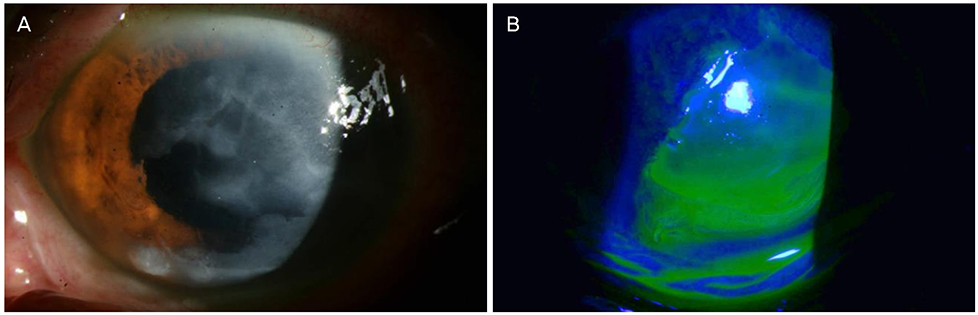
Figure 3 Slit-lamp photograph of the left eye at hospital day 4. (A) Diffuse corneal opacity and keratoprecipitates. (B) Increased corneal epithelial defects in the fluorescein staining were seen.

Figure 4 Slit-lamp photograph of the left eye after 2 months of treatment. (A, B) Fully healed corneal epithelium with remained mild corneal opacity were observed.
Reference
-
1. Ruoff KL, Toutain-Kidd CM, Srinivasan M, et al. Corynebacterium macginleyi isolated from a corneal ulcer. Infect Dis Rep. 2010; 2:1568.
Article2. Riegel P, Ruimy R, de Briel D, et al. Genomic diversity and phylogenetic relationships among lipid-requiring diphtheroids from humans and characterization of Corynebacterium macginleyi sp. nov. Int J Syst Bacteriol. 1995; 45:128–133.
Article3. Suzuki T, lihara H, Uno T, et al. Suture-related keratitis caused by Corynebacterium macginleyi. J Clin Microbiol. 2007; 45:3833–3836.
Article4. Ferrer C, Ruiz-Moreno JM, Rodríguez A, et al. Postoperative Corynebacterium macginleyi endophthalmitis. J Cataract Refract Surg. 2004; 30:2441–2444.
Article5. Funke G, Pagano-Niederer M, Bernauer W. Corynebacterium macginleyi has to date been isolated exclusively from conjunctival swabs. J Clin Microbiol. 1998; 36:3670–3673.
Article6. Espínola M, Somodevilla A, Domingo D, et al. Antibiotic susceptibility of Corynebacterium macginleyi strains causing conjunctivitis. Rev Esp Quimioter. 2010; 23:196–200.7. Joussen AM, Funke G, Joussen F, Herbertz G. Corynebacterium macginleyi: a conjunctiva specific pathogen. Br J Ophthalmol. 2000; 84:1420–1422.
Article8. Dias M, Shreevidya K, Rao SD, Shet D. Corynebacterium macginleyi a rare bacteria causing infection in an immunocompromised patient. J Cancer Res Ther. 2010; 6:374–375.
Article9. Cacopardo B, Stefani S, Cardì F, et al. Surgical site infection by Corynebacterium macginleyi in a patient with neurofibromatosis Type 1. Case Rep Infect Dis. 2013; 970678.10. Kebbe J, Mador MJ. Corynebacterium macginleyi: a cause of ventilator associated pneumonia in an immunocompromised patient. Respir Med Case Rep. 2015; 16:154–156.
Article11. Alsuwaidi AR, Wiebe D, Burdz T, et al. Corynebacterium macginleyi conjunctivitis in Canada. J Clin Microbiol. 2010; 48:3788–3790.
Article12. Bezza Benkaouha I, Le Brun C, Pisella PJ, et al. Bacterial flora in blepharitis. J Fr Ophtalmol. 2015; 38:723–728.13. Eguchi H, Kuwahara T, Miyamoto T, et al. High-level fluoroquinolone resistance in ophthalmic clinical isolates belonging to the species Corynebacterium macginleyi. J Clin Microbiol. 2008; 46:527–532.
Article
- Full Text Links
-
- Actions
-
Cited
- CITED
-
- Close
- Share
-
- Similar articles
-
- Pneumonia Caused by Corynebacterium macginleyi in HIV-infected Patient
- A supplemental treatment for the corneal ulcer or the other corneal diseases
- A Case of Corneal Ulcer Caused by Paecilomyces in Diabetic Patient Wearing Soft Contact Lens
- Treatment of Chronic Corneal Ulcer with Nd-YAG Laser
- Five-Layered Reinforcing Amniotic Membrane Transplantation for Treatment of Deep Corneal Ulcer or Perforation
