Meibomian Gland Dysfunction and Tear Lipid Layer Analysis after Cataract Surgery
- Affiliations
-
- 1Department of Ophthalmology and Visual Science, Yeouido St. Mary's Hospital, College of Medicine, The Catholic University of Korea, Seoul, Korea. sara514@catholic.ac.kr
- KMID: 2449704
- DOI: http://doi.org/10.3341/jkos.2019.60.6.519
Abstract
- PURPOSE
We report the clinical manifestations of dry eye syndrome after cataract surgery involving meibomian gland structure, meibomian gland function, and tear lipid layer analysis.
METHODS
The clinical manifestations of dry eye syndrome were retrospectively evaluated in 34 eyes of 31 patients who underwent cataract surgery from September to November 2017. The ocular surface disease index (OSDI), tear break-up time (tBUT), Oxford stain score, presence or absence of blepharitis, and meibomian gland expression were measured preoperatively and at 1 week, 1 month, and 2 months postoperatively. Lipid layer thickness (LLT), partial blinks, and meibomian gland images were measured using LipiView® (TearScience, Morrisville, NC, USA), an interferometric eye surface measuring device.
RESULTS
The postoperative OSDI was significantly higher than preoperative OSDI (17.09 ± 1.81): 22.76 ± 1.99 at 1 week, 23.12 ± 1.91 at 1 month, and 22.68 ± 1.92 at 2 months (p < 0.05). The postoperative tBUT was significantly lower than preoperative tBUT (5.07 ± 0.39): 3.99 ± 0.31 at 1 week, 3.49 ± 0.27 at 1 month, and 4.72 ± 0.39 at 2 months (p < 0.05). The Oxford staining score increased after surgery, but the difference was not statistically significant. Postoperative meibomian gland expression was significantly lower preoperative values (4.9 ± 2.8): 4.4 ± 2.8 at 1 month, and 3.9 ± 2.8 at 2 months (p < 0.05). The LLT decreased at 1 month postoperatively and increased at 2 months postoperatively, but these differences were not statistically significant.
CONCLUSIONS
Cataract surgery resulted in a short-term meibomian gland dysfunction, leading to deterioration of dry eye after cataract surgery. However, we could not confirm structural changes in the meibomian gland, so it will be necessary to observe the clinical features of dry eye syndrome over a longer period of time.
Figure
-

Figure 1 Images of Meibomian gland drop out, tortuosity, ectasis cases of meibographs, captured by LipiView® (TearScience, Morrisville, NC, USA). Meibography image analysis examples. The freehand tool in ImageJ was used to select the area of meibomian gland, and computerized analysis of the area of Meibomian gland. (A) Meibomian gland drop out, partial or total gland loss or atrophy. (B) Meibomian gland tortuosity. (C) Meibomian gland ectasis, partial or total gland dilatation.
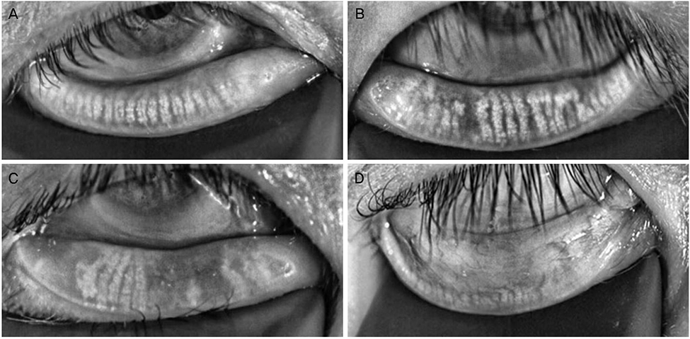
Figure 2 The grading of Meibomian gland loss. Partial or complete loss of Meibomian gland is scored for each eyelid from grade 0 to grade 3. (A) Grade 0 means no loss of meibomian glands, normal Meibomian gland. (B) Grade 1 means the lost area was less than 1/3 of total area. (C) Grade 2 means the lost area was between 1/3 and 2/3 of total area. (D) Grade 3 means the lost area was more than 2/3 of total area.
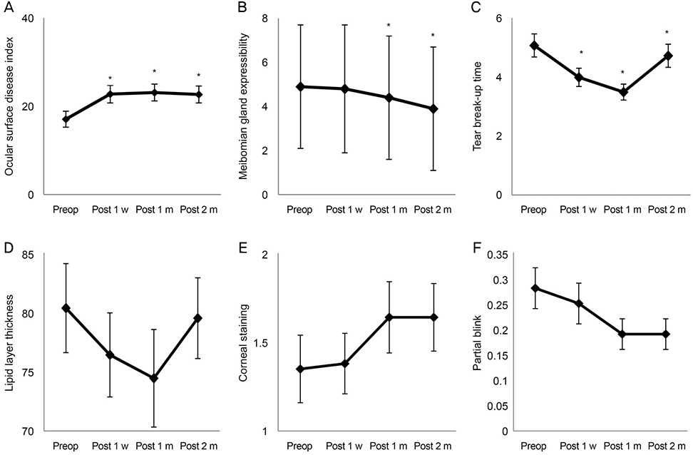
Figure 3 Analysis of the Meibomian gland expressibility, lipid layer thickness, Ocular surface disease index (OSDI), tear film break-up time preop and postop. (A) Change in OSDI from preoperative value. The increase in OSDI was statistically significant at 1 week, 1 and 2 months postoperatively compared to preoperative value (p < 0.05). (B) Change in tear break-up time (tBUT) from preoperative value. tBUT was significantly short at 1 week, 1 and 2 months postoperatively compared to the preoperative value (p < 0.05). (C) Change in corneal staining score from preoperative value. There was an increase in the corneal staining score at 1 week, 1 and 2 months postoperatively (p > 0.05). (D) Change in Meibomian gland expressibility from preoperative value. Meibomian gland expressibility was significantly decreased at 1 and 2 months postoperatively compared to the preoperative value (p < 0.05). (E) Change in lipid layer thickness from preoperative value (p > 0.05). (F) Change in partial blink from preoperative value (p > 0.05). Repeated-measures analysis of variance, compared with preoperative values. Preop = preoperative; Postop = postoperative; w = week(s); m = month(s). *Significant correlations (p < 0.05).
Reference
-
1. Lee HS. The factors influencing health-related quality of life in the elderly: focused on the general characteristics, health habits, mental health, chronic diseases, and nutrient intake status: data from the Fifth Korea National Health and Nutrition Examination Survey (KNHANES V), 2010–2012. Korean J Community Nutr. 2014; 19:479–489.
Article2. Ridder WH III, Oquindo C, Dhamdhere K, Burke J. Does age influence the effect of povidone iodine 5% on the cornea? Austin J Clin Ophthalmol. 2017; 4:1081.3. Moon H, Yoon JH, Hyun SH, Kim KH. Short-term influence of aspirating speculum use on dry eye after cataract surgery: a prospective study. Cornea. 2014; 33:373–375.4. Xu KP, Yagi Y, Tsubota K. Decrease in corneal sensitivity and change in tear function in dry eye. Cornea. 1996; 15:235–239.
Article5. Hwang HB, Kim HS. Phototoxic effects of an operating microscope on the ocular surface and tear film. Cornea. 2014; 33:82–90.
Article6. Massingale ML, Li X, Vallabhajosyula M, et al. Analysis of inflammatory cytokines in the tears of dry eye patients. Cornea. 2009; 28:1023–1027.
Article7. Nwaji ECS, Barrhah GHO. The effect of local anesthetics on tear production. J Nigerian Optom Assoc. 2005; 12:27–29.
Article8. Simone JN, Whitacre MM. Effects of anti-inflammatory drugs following cataract extraction. Curr Opin Ophthalmol. 2001; 12:63–67.
Article9. Choi SH, Kim NJ, Jung SY, Cha MB. Lipid layer thickness in precorneal tear film. J Korean Ophthalmol Soc. 1997; 38:195–200.10. Kim EC. Diagnosis and treatment of dry eye syndrome. J Korean Med Assoc. 2018; 61:352–364.
Article11. Kim JS, Lee H, Choi S, et al. Assessment of the tear film lipid layer thickness after cataract surgery. Semin Ophthalmol. 2018; 33:231–236.
Article12. Miller KL, Walt JG, Mink DR, et al. Minimal clinically important difference for the ocular surface disease index. Arch Ophthalmol. 2010; 128:94–101.
Article13. Bron AJ, Evans VE, Smith JA. Grading of corneal and conjunctival staining in the context of other dry eye tests. Cornea. 2003; 22:640–650.
Article14. Shimazaki J, Sakata M, Tsubota K. Ocular surface changes and discomfort in patients with meibomian gland dysfunction. Arch Ophthalmol. 1995; 113:1266–1270.
Article15. Pult H, Nichols JJ. A review of meibography. Optom Vis Sci. 2012; 89:E760–E769.
Article16. Lam SM, Tong L, Duan X, et al. Longitudinal changes in tear fluid lipidome brought about by eyelid-warming treatment in a cohort of meibomian gland dysfunction. J Lipid Res. 2014; 55:1959–1969.17. Lee JA, Cho YK. The Influence of preoperative meibomian gland disease on dryness after cataract surgery. J Korean Ophthalmol Soc. 2016; 57:228–235.
Article18. Park Y, Hwang HB, Kim HS. Observation of influence of cataract surgery on the ocular surface. PLoS One. 2016; 11:e0152460.
Article19. Smith J, Nichols KK, Baldwin EK. Current patterns in the use of diagnostic tests in dry eye evaluation. Cornea. 2008; 27:656–662.
Article20. Oh T, Jung Y, Chang D, et al. Changes in the tear film and ocular surface after cataract surgery. Jpn J Ophthalmol. 2012; 56:113–118.
Article21. Sahu PK, Das GK, Malik A, Biakthangi L. Dry eye following phacoemulsification surgery and its relation to associated intraoperative risk factors. Middle East Afr J Ophthalmol. 2015; 22:472–477.
Article22. Foulks GN, Bron AJ. Meibomian gland dysfunction: a clinical scheme for description, diagnosis, classification, and grading. Ocul Surf. 2003; 1:107–126.
Article23. Himebaugh NL, Begley CG, Bradley A, Wilkinson JA. Blinking and tear break-up during four visual tasks. Optom Vis Sci. 2009; 86:E106–E114.
Article
- Full Text Links
-
- Actions
-
Cited
- CITED
-
- Close
- Share
-
- Similar articles
-
- The Effects of Warm Compression on Eyelid Temperature and Lipid Layer Thickness of Tear Film
- Efficacy of Intense Pulsed Light Treatment in Patients with Sjögren’s Syndrome Associated with Meibomian Gland Dysfunction
- Investigation of Tear Lipid Layer Patterns According to Blinking Among Dry Eye Patients
- Change of Lipid Layer in Tear Film after Cataract Surgery
- The Influence of Preoperative Meibomian Gland Disease on Dryness after Cataract Surgery
