Diagnosis of Pulmonary Arterial Hypertension in Children by Using Cardiac Computed Tomography
- Affiliations
-
- 1Department of Radiology and Medical Imaging, National Taiwan University Hospital, Medical College, National Taiwan University, Taipei, Taiwan.
- 2Department of Pediatrics, National Taiwan University Hospital, Medical College, National Taiwan University, Taipei, Taiwan. mingtailin@ntu.edu.tw
- 3Department of Surgery, National Taiwan University Hospital, Medical College, National Taiwan University, Taipei, Taiwan.
- KMID: 2447076
- DOI: http://doi.org/10.3348/kjr.2018.0673
Abstract
OBJECTIVE
To establish diagnostic criteria for pulmonary arterial hypertension (PAH) in children by using parameters obtained through noninvasive cardiac computed tomography (CCT).
MATERIALS AND METHODS
We retrospectively measured parameters from CCT images of children from a single institution in a multiple stepwise process. A total of 208 children with mean age of 10.5 years (range: 4 days-18.9 years) were assessed. The variables were classified into three groups: the great arteries; the ventricular walls; and the bilateral ventricular cavities. The relationship between the parameters obtained from the CCT images and mean pulmonary arterial pressure (mPAP) was tested and adjusted by the children's body size. Reference curves for the pulmonary trunk diameter (PTD) and ratio of diameter of pulmonary trunk to ascending aorta (rPTAo) of children with CCT images of normal hearts, adjusted for height, were plotted. Threshold lines were established on the reference curves.
RESULTS
PTD and rPTAo on the CCT images were significantly positively correlated with mPAP (r > 0.85, p < 0.01). Height was the body size parameter most correlated with PTD (r = 0.91, p < 0.01) and rPTAo (r = −0.69, p < 0.01). On the basis of the threshold lines on the reference curves, PTD and rPTAo both showed 88.9% sensitivity for PAH diagnosis, with negative predictive values of 93.3% and 92.9%, respectively.
CONCLUSION
PTD and rPTAo measured from CCT images were significantly correlated with mPAP in children. Reference curves and the formula of PTD and rPTAo adjusted for height could be practical for diagnosing PAH in children.
MeSH Terms
Figure
-
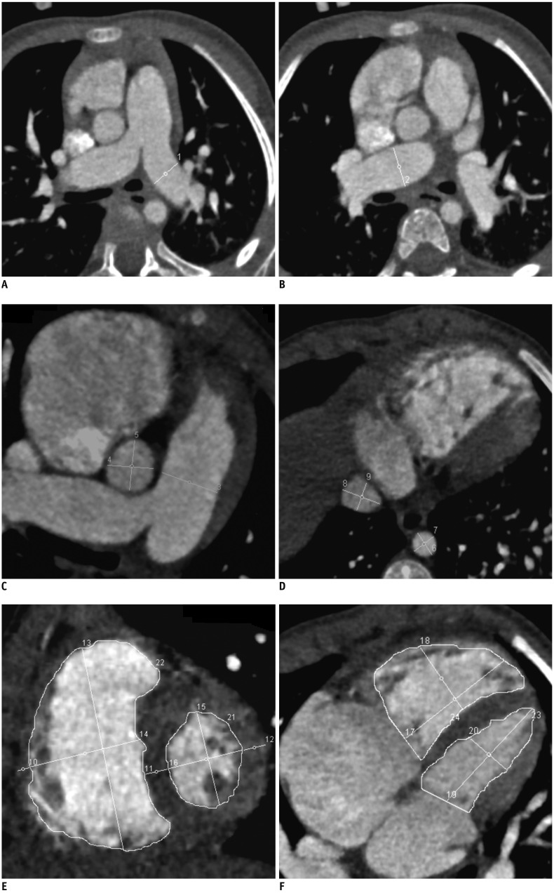
Fig. 1 Parameters measured on end-diastole phase of contrast-enhanced CCT images to diagnose pulmonary arterial hypertension.A, B, D. Images were obtained in transverse planes. C. Image was obtained in tilted oblique axial plane to reveal whole pulmonary trunk from pulmonary annulus to bifurcation. E, F. Images were at middle ventricular level with image (E) on cardiac short-axis plane and image (F) on cardiac four-chamber plane. Twenty measurements were performed and annotated 1–20, and all of these measurements could also be easily performed on picture archiving and communication system. 1 = maximal diameter of left pulmonary artery before branching, 2 = maximal diameter of right pulmonary artery before branching, 3 = maximal diameter of middle pulmonary trunk before bifurcation, 4 and 5 = perpendicular diameters of ascending aorta on same image that measured pulmonary trunk, 6 and 7 = perpendicular diameters of descending aorta at level through diaphragm, 8 and 9 = perpendicular diameters of inferior vena cava on same image that measured descending aorta, 10–12 = middle ventricular myocardial thickness of RV, septum, and LV respectively, 13 and 14, 15 and 16 = perpendicular height and width intersects at midpoints of RV and LV, respectively, 17 and 18, 19 and 20 = perpendicular length and width intersects at midpoints of RV and LV respectively, 21 and 23, 22 and 24 = area of LV and RV respectively. CCT = cardiac computed tomography, LV = left ventricle, RV = right ventricle
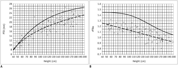
Fig. 2 Relationship of PTD (A) and rPTAo (B) to height in children determined using CCT images of normal hearts.Dashes represent best-fitting linear regression of mean of study subjects from our study step II. Solid lines indicate thresholds of 25-mm Hg mean pulmonary arterial pressure calculated from subjects from our study step III. Subjects from steps II and III were not same. PTD = pulmonary trunk diameter, rPTAo = PTD-to-ascending aorta diameter ratio
Reference
-
1. Humbert M, Sitbon O, Yaïci A, Montani D, O'Callaghan DS, Jaïs X, et al. French Pulmonary Arterial Hypertension Network. French Pulmonary Arterial Hypertension Network. Survival in incident and prevalent cohorts of patients with pulmonary arterial hypertension. Eur Respir J. 2010; 36:549–555. PMID: 20562126.2. Fowler NO, Westcott RN, Scott RC. Normal pressure in right heart and pulmonary artery. Am Heart J. 1953; 46:264–267. PMID: 13080163.3. Peacock AJ, Murphy NF, McMurray JJ, Caballero L, Stewart S. An epidemiological study of pulmonary arterial hypertension. Eur Respir J. 2007; 30:104–109. PMID: 17360728.
Article4. Gupta SK, Saxena A, Gulati GS. Evaluation of pulmonary hypertension in a child: role of computed tomography. Indian J Pediatr. 2011; 78:1417–1419. PMID: 21625838.
Article5. Nauser TD, Stites SW. Diagnosis and treatment of pulmonary hypertension. Am Fam Physician. 2001; 63:1789–1798. PMID: 11352291.6. Celermajer DS, Marwick T. Echocardiographic and right heart catheterization techniques in patients with pulmonary arterial hypertension. Int J Cardiol. 2008; 125:294–303. PMID: 17689753.
Article7. Schannwell CM, Steiner S, Strauer BE. Diagnostics in pulmonary hypertension. J Physiol Pharmacol. 2007; 58(Suppl 5):(Pt 2):591–602.8. Hegewald MJ, Markewitz B, Elliott CG. Pulmonary hypertension: clinical manifestations, classification and diagnosis. Int J Clin Pract Suppl. 2007; (156):5–14.
Article9. McLaughlin VV, McGoon MD. Primary pulmonary hypertension. Hospital Physician Board Review Manual. 2002; 9:1–11.10. Bossone E, Duong-Wagner TH, Paciocco G, Oral H, Ricciardi M, Bach DS, et al. Echocardiographic features of primary pulmonary hypertension. J Am Soc Echocardiogr. 1999; 12:655–662. PMID: 10441222.
Article11. McGoon M, Gutterman D, Steen V, Barst R, McCrory DC, Fortin TA, et al. Screening, early detection, and diagnosis of pulmonary arterial hypertension: ACCP evidence-based clinical practice guidelines. Chest. 2004; 126(1 Suppl):14S–34S. PMID: 15249493.12. ASCI CCT & CMR Guideline Working Group. Tsai IC, Choi BW, Chan C, Jinzaki M, Kitagawa K, et al. ASCI 2010 appropriateness criteria for cardiac computed tomography: a report of the Asian Society of Cardiovascular Imaging Cardiac Computed Tomography and Cardiac Magnetic Resonance Imaging Guideline Working Group. Int J Cardiovasc Imaging. 2010; 26(Suppl 1):1–15.
Article13. Tsai IC, Goo HW. Cardiac CT and MRI for congenital heart disease in Asian countries: recent trends in publication based on a scientific database. Int J Cardiovasc Imaging. 2013; 29(Suppl 1):1–5. PMID: 23344910.
Article14. Yang JC, Lin MT, Jaw FS, Chen SJ, Wang JK, Shih TT, et al. Trends in the utilization of computed tomography and cardiac catheterization among children with congenital heart disease. J Formos Med Assoc. 2015; 114:1061–1068. PMID: 25241602.
Article15. Kuriyama K, Gamsu G, Stern RG, Cann CE, Herfkens RJ, Brundage BH. CT-determined pulmonary artery diameters in predicting pulmonary hypertension. Invest Radiol. 1984; 19:16–22. PMID: 6706516.
Article16. Ley S, Kreitner KF, Fink C, Heussel CP, Borst MM, Kauczor HU. Assessment of pulmonary hypertension by CT and MR imaging. Eur Radiol. 2004; 14:359–368. PMID: 14740163.
Article17. Di Guglielmo L, Dore R, Vespro V. Pulmonary hypertension: role of computed tomography and magnetic resonance imaging. Ital Heart J. 2005; 6:846–851. PMID: 16270478.18. McLure LE, Peacock AJ. Imaging of the heart in pulmonary hypertension. Int J Clin Pract. 2007; 61(S156):15–26. PMID: 17229176.
Article19. Chen BB, Chen SJ, Wu MH, Li YW, Lue HC. EBCT-McGoon ratio a reliable and useful method to predict pulmonary blood flow non-invasively. Chinese J Radiol. 2007; 32:1–8.20. Goo HW. State-of-the-art CT imaging techniques for congenital heart disease. Korean J Radiol. 2010; 11:4–18. PMID: 20046490.
Article21. Corson N, Armato SG 3rd, Labby ZE, Straus C, Starkey A, Gomberg-Maitland M. CT-based pulmonary artery measurements for the assessment of pulmonary hypertension. Acad Radiol. 2014; 21:523–530. PMID: 24594422.
Article22. Ng CS, Wells AU, Padley SP. A CT sign of chronic pulmonary arterial hypertension: the ratio of main pulmonary artery to aortic diameter. J Thorac Imaging. 1999; 14:270–278. PMID: 10524808.
Article23. Haimovici JB, Trotman-Dickenson B, Halpern EF, Dec GW, Ginns LC, Shepard JA, et al. Relationship between pulmonary artery diameter at computed tomography and pulmonary artery pressures at right-sided heart catheterization. Massachusetts General Hospital lung transplantation program. Acad Radiol. 1997; 4:327–334. PMID: 9156228.24. Mullen MP. Diagnostic strategies for acute presentation of pulmonary hypertension in children: particular focus on use of echocardiography, cardiac catheterization, magnetic resonance imaging, chest computed tomography, and lung biopsy. Pediatr Crit Care Med. 2010; 11(2 Suppl):S23–S26. PMID: 20216157.
Article25. Lee YH, Song GG. Meta-analysis of randomized controlled trials of bosentan for treatment of pulmonary arterial hypertension. Korean J Intern Med. 2013; 28:701–707. PMID: 24307846.
Article26. Wang RC, Jiang FM, Zheng QL, Li CT, Peng XY, He CY, et al. Efficacy and safety of sildenafil treatment in pulmonary arterial hypertension: a systematic review. Respir Med. 2014; 108:531–537. PMID: 24462476.
Article27. Jin KN, Park EA, Shin CI, Lee W, Chung JW, Park JH. Retrospective versus prospective ECG-gated dual-source CT in pediatric patients with congenital heart diseases: comparison of image quality and radiation dose. Int J Cardiovasc Imaging. 2010; 26(Suppl 1):63–73. PMID: 20044793.
Article28. Goo HW. CT radiation dose optimization and estimation: an update for radiologists. Korean J Radiol. 2012; 13:1–11. PMID: 22247630.
Article29. Remy-Jardin M, Delhaye D, Teisseire A, Hossein-Foucher C, Duhamel A, Remy J. MDCT of right ventricular function: impact of methodologic approach in estimation of right ventricular ejection fraction, part 2. AJR Am J Roentgenol. 2006; 187:1605–1609. PMID: 17114557.
Article30. Savino G, Zwerner P, Herzog C, Politi M, Bonomo L, Costello P, et al. CT of cardiac function. J Thorac Imaging. 2007; 22:86–100. PMID: 17325580.
Article31. Nakamura K, Miyahara Y, Ikeda S, Naito T. Assessment of right ventricular diastolic function by pulsed Doppler echocardiography in chronic pulmonary disease and pulmonary thromboembolism. Respiration. 1995; 62:237–243. PMID: 8560088.
Article32. Bugnone AN, Viamonte M Jr, Garcia H. Imaging findings in human immunodeficiency virus-related pulmonary hypertension: report of five cases and review of the literature. Radiology. 2002; 223:820–827. PMID: 12034955.
Article
- Full Text Links
-
- Actions
-
Cited
- CITED
-
- Close
- Share
-
- Similar articles
-
- Pulmonary Arterial Hypertension with Congenital Heart Diseases
- Updated clinical classification of pulmonary hypertension
- Diagnosis and treatment of idiopathic pulmonary arterial hypertension
- Pulmonary Arterial Hypertension
- The effect of perioperative inhaled iloprost on congenital heart disease with severe pulmonary arterial hypertension
