Mucinous Non-neoplastic Cyst of the Pancreas
- Affiliations
-
- 1Department of Surgery, Wonkwang University School of Medicine, Iksan, Korea. knife@wonkwang.ac.kr
- 2Department of Pathology, Wonkwang University School of Medicine, Iksan, Korea.
- KMID: 2443638
- DOI: http://doi.org/10.4166/kjg.2019.73.4.235
Abstract
- Cystic neoplasms of the pancreas consist of a wide range of pathological entities and are being detected more frequently due to advances in cross-sectional imaging modalities and increasing numbers of periodic health checkups. The majority of pancreatic cystic neoplasms are intraductal papillary mucinous neoplasms, serous neoplasms, and mucinous cystic neoplasms, but recently, rare cases of mucinous non-neoplastic cyst of the pancreas (MNCP) have been reported, and despite the availabilities of modern imaging systems, such as MRI and CT, the differentiation of non-neoplastic and neoplastic cysts remains challenging. Herein, we report our experience of a 65-year-old male case with an MNCP.
Keyword
MeSH Terms
Figure
-
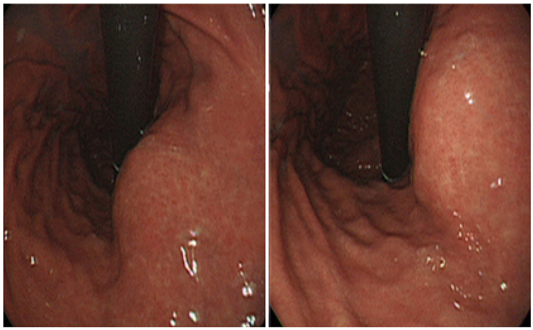
Fig. 1 Upper gastrointestinal endoscopy revealed an external compressive lesion at the posterior wall of the stomach midbody.

Fig. 2 (A) Abdominal computed tomography image showing a 2.5 cm, round, hypoattenuated cystic lesion without any enhanced solid portion in the pancreatic body. (B) Three-dimensional magnetic resonance cholangiopancreatogram showing a 2.5 cm, cystic mass without any solid portion in the pancreatic body surrounded by multiple, small daughter cysts. (C) Endoscopic ultrasound revealed a non-septated, 2.5 cm, single cyst with mural nodularity. Circles and an arrow indicated a cystic lesion.

Fig. 3 (A) Cut surface of the resected specimen showing the unilocular cyst. (B) H&E-stained section shows the cyst lined with mucinous epithelial cells (arrow), a fibrous stroma (arrowhead) and adjacent normal pancreas (asterisk) (H&E, ×100).
Reference
-
1. Kosmahl M, Egawa N, Schröder S, Carneiro F, Lüttges J, Klöppel G. Mucinous nonneoplastic cyst of the pancreas: a novel nonneoplastic cystic change? Mod Pathol. 2002; 15:154–158.
Article2. Klöppel G, Kosmahl M. Cystic lesions and neoplasms of the pancreas. The features are becoming clearer. Pancreatology. 2001; 1:648–655.3. Habashi S, Draganov PV. Pancreatic pseudocyst. World J Gastroenterol. 2009; 15:38–47.
Article4. Cao W, Adley BP, Liao J, et al. Mucinous nonneoplastic cyst of the pancreas: apomucin phenotype distinguishes this entity from intraductal papillary mucinous neoplasm. Hum Pathol. 2010; 41:513–521.
Article5. Kosmahl M, Pauser U, Peters K, et al. Cystic neoplasms of the pancreasand tumor-like lesions with cystic features: a review of 418 cases and a classification proposal. Virchows Arch. 2004; 445:168–178.
Article6. Yang JD, Song JS, Noh SJ, Moon WS. Mucinous non-neoplastic cyst of the pancreas. Korean J pathol. 2013; 47:188–190.
Article7. Brugge WR, Lauwers GY, Sahani D, Fernandez-del Castillo C, Warshaw AL. Cystic neoplasms of the pancreas. N Engl J Med. 2004; 351:1218–1226.
Article8. Nagula S, Kennedy T, Schattner MA, et al. Evaluation of cyst fluid CEA analysis in the diagnosis of mucinous cysts of the pancreas. J Gastrointest Surg. 2010; 14:1997–2003.
Article9. Lewandrowski KB, Southern JF, Pins MR, Compton CC, Warshaw AL. Cyst fluid analysis in the differential diagnosis of pancreatic cysts. A comparison of pseudocysts, serous cystadenomas, mucinous cystic neoplasms, and mucinous cystadenocarcinoma. Ann Surg. 1993; 217:41–47.
Article
