Intramural Gastric Hematoma after Acute Necrotizing Pancreatitis: A Case Report and Review of Imaging Findings
- Affiliations
-
- 1Department of Radiology, Gil Medical Center, Gachon University College of Medicine, Incheon, Korea. nnoleeter@naver.com
- 2Department of Internal medicine, Gil Medical Center, Gachon University College of Medicine, Incheon, Korea.
- KMID: 2442470
- DOI: http://doi.org/10.3348/jksr.2019.80.1.117
Abstract
- Intramural hematoma of the gastrointestinal tract is a rare disease entity. Pancreatitis-induced intramural gastric hematoma (IGH) is far more seldom reported. Here, we report a rare case of a giant IGH occurring as a delayed complication of pancreatitis in a 51-year-old man. The diagnosis was made using computed tomography (CT) and endoscopic ultrasonography. The patient was conservatively managed, and follow-up abdominal CT showed marked decreases in the size of the IGH.
MeSH Terms
Figure
-
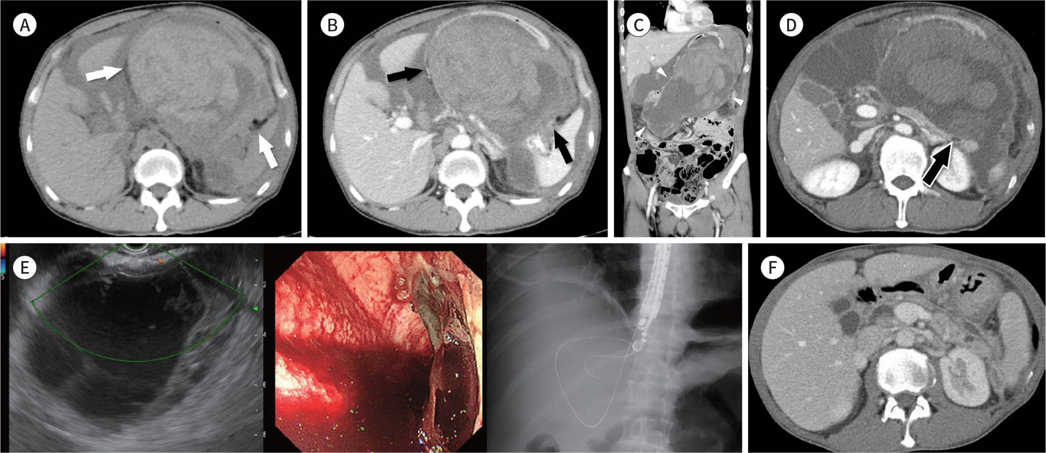
Fig. 1. A 51-year-old man with an IGH associated with pancreatitis. A–C. Axial CT scans showing a mixture of fluid-dense and hyperdense lesions (50–70 Hounsfield units) (A) without enhancement (white arrows) and (B) along the greater curvature of the stomach (black arrows). (C) Coronal CT scan showing a huge hematoma (white arrowheads) along the gastric wall and distension of the gastric wall. There is a small amount of fluid collection with mild peritoneal thickening in the omentum and perisplenic space, suggesting remnant or recurrent walled-off necrosis of pancreatitis. D. A small tubular structure (arrow) between the pancreatic tail and IGH, suggesting the possible presence of a pancreaticogastric fistula. E. Endoscopic ultrasonography shows a large cystic lesion with heterogeneous echogenicity and internal septation in the greater curvature of the gastric wall. There was no vascularity within the mass. About 200 mL of fluid was aspirated. As the fluid appeared dark reddish in color,the mass was confirmed as a hematoma. F. Follow-up abdominal CT scan obtained after about 2 months showing a marked decrease in the size of IGH, and walled-off necrosis around the pancreas tail and retroperitoneum.
Reference
-
References
1. Imaizumi H, Mitsuhashi T, Hirata M, Aizaki T, Nishimaki H, Soma K, et al. A giant intramural gastric hematoma successfully treated by transcatheter arterial embolization. Intern Med. 2000; 39:231–234.
Article2. Dhawan V, Mohamed A, Fedorak RN. Gastric intramural hematoma: a case report and literature review. Can J Gastroenterol. 2009; 23:19–22.
Article3. Cimavilla Román M, Torres Yuste R, Vallecillo Sande MA. Intramural gastric hematoma in the context of an acute pancreatitis. Rev Esp Enferm Dig. 2017; 109:170.
Article4. Chou CT, Chen RC, Yang AD. Gastric subserosal hematoma developing from focal pancreatitis: a case report. Kaohsiung J Med Sci. 2009; 25:45–48.
Article5. Bailey WC, Akers DR. Traumatic intramural hematoma of the duodenum in children. A report of five cases. Am J Surg. 1965; 110:695–703.6. Fielding GA, McLatchie GR, Wilson C, Imrie CW, Carter DC. Acute pancreatitis and pancreatic fistula formation. Br J Surg. 1989; 76:1126–1128.
Article7. van Spreeuwel JP, van Gorp LH, Bast TJ, Nadorp JH. Intramural hematoma of the duodenum in a patient with chronic pancreatitis. Endoscopy. 1981; 13:246–248.
Article8. Ma JK, Ng KK, Poon RT, Fan ST. Pancreatic-induced intramural duodenal haematoma. Asian J Surg. 2008; 31:83–86.
Article
- Full Text Links
-
- Actions
-
Cited
- CITED
-
- Close
- Share
-
- Similar articles
-
- Successful Endoscopic Decompression for Intramural Duodenal Hematoma with Gastric Outlet Obstruction Complicating Acute Pancreatitis
- Intramural Duodenal Hematoma after Transpancreatic Septotomy during ERCP: A Case Report and Literature Review
- Acute Pancreatitis and Gastroduodenal Intussusception Induced by an Underlying Gastric Gastrointestinal Stromal Tumor: A Case Report
- Intramural Duodenal Hematoma Caused by Acute Gallstone Pancreatitis
- A Case of Duodenal Intramural Hematoma Caused by Acute Alcoholic Pancreatitis
