Anat Cell Biol.
2019 Mar;52(1):90-92. 10.5115/acb.2019.52.1.90.
Clinical importance of tensor fasciae suralis arising from linea aspera along with short head of biceps femoris: a rare anomaly
- Affiliations
-
- 1Department of Anatomy, Melaka Manipal Medical College (Manipal Campus), Manipal Academy of Higher Education, Manipal, India. nayaksathish@gmail.com
- KMID: 2442336
- DOI: http://doi.org/10.5115/acb.2019.52.1.90
Abstract
- Tensor fasciae suralis, also known as ischioaponeuroticus is a clinically relevant muscle variant located in the popliteal fossa. Though rare, when present the muscle may arise from any of the hamstrings and gets inserted to the crural fascia of leg or tendocalcaneus and is innervated by the tibial component of sciatic nerve. Here we report a variant of tensor fasciae suralis originated from the lowermost part of linea aspera along with the fibers of short head of biceps femoris in the left lower limb of a male cadaver aged approximately 58 years. The muscle was 16 cm in length and 1 cm breadth in its widest part. It was found inserted to the crural fascia over the lateral head of gastrocnemius and was found innervated by common peroneal nerve. To the best of our knowledge, the tensor fascia suralis muscle originated from linea aspera along with short head of biceps femoris and innervated by common peroneal nerve has not been reported in either cadaveric or imaging studies.
Keyword
Figure
-
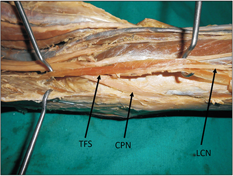
Fig. 1 Dissected popliteal fasciae. Note the tensor fasciae suralis (TFS) overlapping the common peroneal nerve (CPN) and lateral cutaneous nerve of the calf (LCN). Origin of TFS from linea aspera also visible.
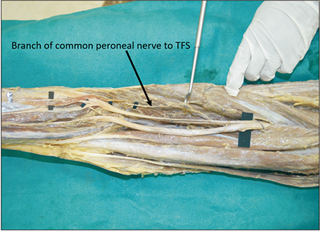
Fig. 2 Distant view of the popliteal fasciae. Note the motor nerve supply to tensor fasciae suralis (TFS) from common peroneal nerve.
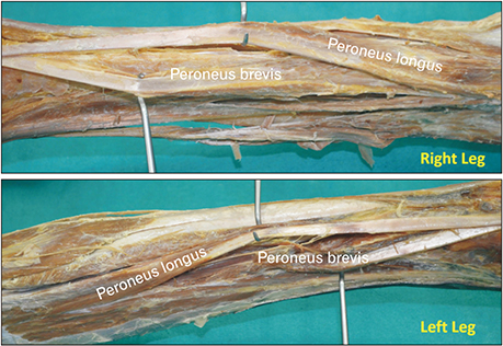
Fig. 3 A composite photograph showing both right and left legs to compare the peroneus longus and brevis muscles of two sides. The peroneal muscles were about 3 mm broader in the right leg when compared to the left leg.
Reference
-
1. Kumar GR, Bhagwat SS. An anomalous muscle in the region of the popliteal fossa: a case report. J Anat Soc India. 2006; 55:65–68.2. Rajendiran R, Murugesan A. Unilateral tensor fascia suralis: a case report. Brunei Darussalam J Health. 2016; 6:94–98.3. Montet X, Sandoz A, Mauget D, Martinoli C, Bianchi S. Sonographic and MRI appearance of tensor fasciae suralis muscle, an uncommon cause of popliteal swelling. Skeletal Radiol. 2002; 31:536–538.
Article4. Kim KH, Shim JC, Lee GJ, Lee KE, Kim HK, Suh JH. MR imaging and ultrasonographic findings of tensor fasciae suralis muscle: a case report. J Korean Soc Radiol. 2015; 73:249–251.
Article5. Gandhi KR, Wabale RN, Farooqui MS. Bilateral presentation of tensor fascia suralis muscle in a male cadaver. Int J Anat Res. 2015; 3:1745–1748.
- Full Text Links
-
- Actions
-
Cited
- CITED
-
- Close
- Share
-
- Similar articles
-
- MR Imaging and Ultrasonographic Findings of Tensor Fasciae Suralis Muscle: A Case Report
- Anomalous Biceps Femoris Tendon Insertion Leading to a Snapping Knee in a Young Male
- Anatomical and Radiological Study of the Vascular Distribution and Skin Territory for the Tensor Fasciae Latae Free Flap
- Transfer of the Medial Hamstring or Biceps Femoris to the Patella
- Complete Rupture of the Proximal Hamstring
