Dentofacial transverse development in Koreans according to skeletal maturation: A cross-sectional study
- Affiliations
-
- 1Department of Orthodontics, Gangnam Severance Dental Hospital, Institute of Craniofacial Deformity, College of Dentistry, Yonsei University, Seoul, Korea. khkim@yuhs.ac
- 2Department of Orthodontics, Institute of Craniofacial Deformity, College of Dentistry, Yonsei University, Seoul, Korea.
- 3Biostatistics Collaboration Unit, College of Medicine, Yonsei University, Seoul, Korea.
- KMID: 2441440
- DOI: http://doi.org/10.4041/kjod.2018.48.1.39
Abstract
OBJECTIVE
The aim of this study was to establish the normative data of dentofacial transverse dimensions according to the skeletal maturation stage in Korean adolescents with good occlusion, assess gender differences and determine correlations between transverse variables.
METHODS
A total of 577 Korean subjects between ages 7 to 19 years and exhibiting skeletal Class I occlusion were categorized by skeletal maturation index (SMI) of Fishman using hand-wrist radiographs. Dentofacial transverse dimensions were assessed using posteroanterior cephalograms. Independent two-sample t-tests were used to analyze differences between genders. Pearson correlation coefficient was used to determine the correlation between transverse measurements.
RESULTS
Dentofacial transverse norms relevant to skeletal maturation stages were established. The average maxillomandibular width difference and ratio at growth completion was 22.16 mm and 77.01% for males; 23.70 mm and 74.06% for females, respectively. Males had greater facial, maxillary and mandibular widths compared to females at every SMI stage. The maxillary and mandibular intermolar widths showed the strongest correlation for both sexes (r = 0.826 for males, r = 0.725 for females).
CONCLUSIONS
Dentofacial transverse norms of Korean adolescents were established according to developmental stage. All dentofacial widths were greater in males at growth completion. Maxillary and mandibular intermolar widths were strongly correlated. This study may serve as a guideline for the assessment of dentofacial transverse growth according to skeletal maturation stage in Korean adolescents with good occlusion.
Keyword
MeSH Terms
Figure
-
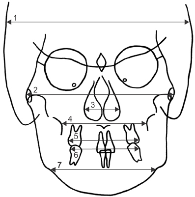
Figure 1 Landmarks and transverse measurements. 1. Cranial width (bieuryon width): distance between the most lateral points on the cranium. 2. Facial width (bizygomatic width): distance between the most lateral points on the zygomatic arch. 3. Nasal width: greatest distance between the right and left lateral bony walls of the nasal cavity. 4. Maxillary width: distance between the right and left jugal process. The jugal process is the intersection of the maxillary tuberosity outline and the zygomatic buttress. 5. Maxillary intermolar width: distance between the most lateral points on the buccal surfaces of the permanent maxillary first molar crowns. 6. Mandibular intermolar width: distance between the most lateral points on the buccal surfaces of the permanent mandibular first molar crowns. 7. Mandibular width: distance between the right and left antegonial notch.
Cited by 2 articles
-
Stability of bimaxillary surgery involving intraoral vertical ramus osteotomy with or without presurgical miniscrew-assisted rapid palatal expansion in adult patients with skeletal Class III malocclusion
Yoon-Soo Ahn, Sung-Hwan Choi, Kee-Joon Lee, Young-Soo Jung, Hyoung-Seon Baik, Hyung-Seog Yu
Korean J Orthod. 2020;50(5):304-313. doi: 10.4041/kjod.2020.50.5.304.Benefits of lateral cephalogram during landmark identification on posteroanterior cephalograms
Sel-Ae Hwang, Jae-Seo Lee, Hyeon-Shik Hwang, Kyung-Min Lee
Korean J Orthod. 2019;49(1):32-40. doi: 10.4041/kjod.2019.49.1.32.
Reference
-
1. Vanarsdall RL Jr. Transverse dimension and long-term stability. Semin Orthod. 1999; 5:171–180.
Article2. Snodell SF, Nanda RS, Currier GF. A longitudinal cephalometric study of transverse and vertical craniofacial growth. Am J Orthod Dentofacial Orthop. 1993; 104:471–483.
Article3. Betts NJ, Vanarsdall RL, Barber HD, Higgins-Barber K, Fonseca RJ. Diagnosis and treatment of transverse maxillary deficiency. Int J Adult Orthodon Orthognath Surg. 1995; 10:75–96.4. Lim HM, Park YC, Kee KJ, Kim KH, Choi YJ. Stability of dental, alveolar, and skeletal changes after miniscrew-assisted rapid palatal expansion. Korean J Orthod. 2017; 47:313–322.
Article5. Park JJ, Park YC, Lee KJ, Cha JY, Tahk JH, Choi YJ. Skeletal and dentoalveolar changes after miniscrew-assisted rapid palatal expansion in young adults: A cone-beam computed tomography study. Korean J Orthod. 2017; 47:77–86.
Article6. Yavuz I, Ikbal A, Baydaş B, Ceylan I. Longitudinal posteroanterior changes in transverse and vertical craniofacial structures between 10 and 14 years of age. Angle Orthod. 2004; 74:624–629.7. Cortella S, Shofer FS, Ghafari J. Transverse development of the jaws: norms for the posteroanterior cephalometric analysis. Am J Orthod Dentofacial Orthop. 1997; 112:519–522.
Article8. Lux CJ, Conradt C, Burden D, Komposch G. Transverse development of the craniofacial skeleton and dentition between 7 and 15 years of age--a longitudinal postero-anterior cephalometric study. Eur J Orthod. 2004; 26:31–42.
Article9. Hägg U, Taranger J. Maturation indicators and the pubertal growth spurt. Am J Orthod. 1982; 82:299–309.
Article10. Hunter CJ. The correlation of facial growth with body height and skeletal maturation at adolescence. Angle Orthod. 1966; 36:44–54.11. Johnston FE, Hufham HP Jr, Moreschi AF, Terry GP. Skeletal maturation and cephalofacial development. Angle Orthod. 1965; 35:1–11.12. Fishman LS. Maturational patterns and prediction during adolescence. Angle Orthod. 1987; 57:178–193.13. Fishman LS. Radiographic evaluation of skeletal maturation. A clinically oriented method based on hand-wrist films. Angle Orthod. 1982; 52:88–112.14. Scammon RE. A summary of the anatomy of the infant and child. In : Abt IA, editor. Pediatrics. Philadelphia: W.B. Saunders;1923. p. 89.15. Ricketts RM. Perspectives in the clinical application of cephalometrics. The first fifty years. Angle Orthod. 1981; 51:115–150.16. Woods GA Jr. Changes in width dimensions between certain teeth and facial points during human growth. Am J Orthod. 1950; 36:676–700.
Article17. Enoki K, Hioki K, Motohashi Y, Kikuchi S, Kwakami N, Kobayashi K, et al. Normal standards for facial growth analysis. J Jap Orthodont Soc. 1958; 17:21–30.18. Wei SH. Craniofacial width dimensions. Angle Orthod. 1970; 40:141–147.19. Edwards CB, Marshall SD, Qian F, Southard KA, Franciscus RG, Southard TE. Longitudinal study of facial skeletal growth completion in 3 dimensions. Am J Orthod Dentofacial Orthop. 2007; 132:762–768.
Article20. Melsen B. Palatal growth studied on human autopsy material. A histologic microradiographic study. Am J Orthod. 1975; 68:42–54.21. Savara BS, Singh IJ. Norms of size and annual increments of seven anatomical measures of maxillae in boys from three to sixteen years of age. Angle Orthod. 1968; 38:104–120.22. Wagner DM, Chung CH. Transverse growth of the maxilla and mandible in untreated girls with low, average, and high MP-SN angles: a longitudinal study. Am J Orthod Dentofacial Orthop. 2005; 128:716–723. quiz 801.
Article23. Athanasiou AE, Droschl H, Bosch C. Data and patterns of transverse dentofacial structure of 6- to 15-year-old children: a posteroanterior cephalometric study. Am J Orthod Dentofacial Orthop. 1992; 101:465–471.
Article24. Gu Y, McNamara JA Jr, Sigler LM, Baccetti T. Comparison of craniofacial characteristics of typical Chinese and Caucasian young adults. Eur J Orthod. 2011; 33:205–211.
Article25. Miyajima K, McNamara JA Jr, Kimura T, Murata S, Iizuka T. Craniofacial structure of Japanese and European-American adults with normal occlusions and well-balanced faces. Am J Orthod Dentofacial Orthop. 1996; 110:431–438.
Article26. Huertas D, Ghafari J. New posteroanterior cephalometric norms: a comparison with craniofacial measures of children treated with palatal expansion. Angle Orthod. 2001; 71:285–292.27. Meredith HV. Growth in bizygomatic face breadth during childhood. Growth. 1954; 18:111–134.28. Baik HS, Yu HS, Lee KJ. A posteroanterior cephalometric study on craniofacial proportions of Koreans with normal occlusion. Korean J Orthod. 1997; 27:643–659.29. Gandini LG Jr, Buschang PH. Maxillary and mandibular width changes studied using metallic implants. Am J Orthod Dentofacial Orthop. 2000; 117:75–80.
Article30. Sawchuk D, Currie K, Vich ML, Palomo JM, Flores-Mir C. Diagnostic methods for assessing maxillary skeletal and dental transverse deficiencies: a systematic review. Korean J Orthod. 2016; 46:331–342.
Article
- Full Text Links
-
- Actions
-
Cited
- CITED
-
- Close
- Share
-
- Similar articles
-
- Mandibular midline osteotomy for correction of bimaxillary transverse discrepancy: a technical note
- Incidence and Skeletal Features of Developmental Cervical and Lumbar Spinal Stenosis
- Assessment of pharyngeal airway in Korean adolescents according to skeletal pattern, sex, and cervical vertebral maturation: A cross-sectional CBCT study
- Assessment of Midpalatal Suture Maturation by Skeletal Maturity on Hand Wrist Radiographs
- Sexual Maturation, Attitudes towards Sexual Maturity, and Body Esteem in Elementary-School Children
