Rapid Spontaneous Resolution of Contralateral Acute Subdural Hemorrhage Caused by Overdrainage of Chronic Subdural Hemorrhage
- Affiliations
-
- 1Department of Neurosurgery, Inje University Haeundae Paik Hospital, Busan, Korea. jheaj@paik.ac.kr
- KMID: 2434838
- DOI: http://doi.org/10.18700/jnc.180051
Abstract
- BACKGROUND
Since the first report of a rapidly resolved subdural hemorrhage (SDH) in 1986, few additional case reports have been presented in the literature.
CASE REPORT
An 82-year-old female patient presented with a SDH over the left convexity. The SDH was removed via catheter drainage through a burr hole trephination. Post-operative computed tomography (CT) following 300 mL drainage from the chronic SDH demonstrated a newly developed SDH along the right convexity. A follow-up CT performed 2 hours later revealed an unexpected significant resolution of the acute SDH.
CONCLUSION
The spontaneous resolution of acute SDH is believed to result from redistribution by washout of the hematoma by cerebrospinal fluid dilution. However, its exact pathophysiology is not well understood. When surgical evacuation is considered in acute SDH, conservative management should also be considered because spontaneous resolution of hemorrhage remains a possibility.
MeSH Terms
Figure
-
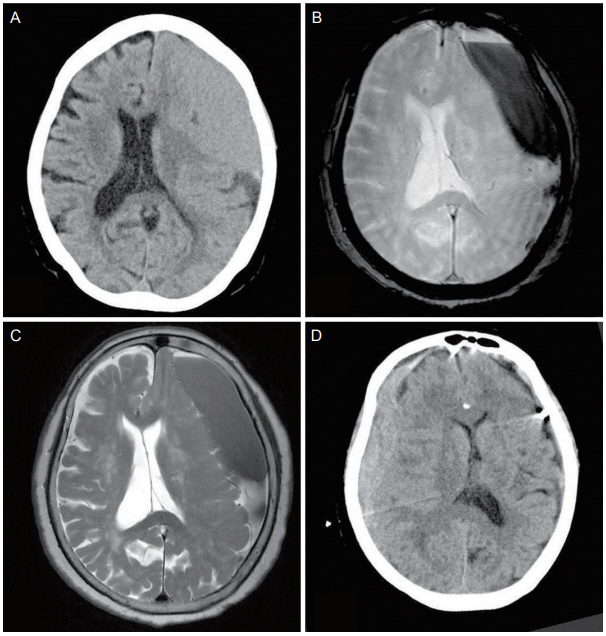
Figure 1. (A) Non-contrast computed tomography demonstrated a SDH over the left convexity. (B) Gradient-echo and (C) T2-weighted images revealing a SDH in the left convexity. (D) Post-operative computed tomography following drainage of the left hematoma, demonstrating a newly developed subdural hemorrhage along the right convexity. SDH, subdural hemorrhage.

Figure 2. (A) A follow-up CT performed 2 hours later revealed spontaneous resolution of the hemorrhage, and a hyperdense band consistent with bleeding (black arrow), in addition to a layered hypodense component consistent with cerebrospinal fluid (white arrow). (B) A CT performed post-operatively on day 2 revealed a further diminished hemorrhage. (C) A CT performed post-operatively on day 4 revealed a further decreased hemorrhage at the right convexity. (D) A CT performed post-operatively on day 7 revealed little hemorrhages at the right convexity. CT, computed tomography.
Reference
-
1. Kim CH, Song GS, Kim YH, Kim YS, Sung SK, Son DW, et al. Remote hemorrhage after burr hole drainage of chronic subdural hematoma. Korean J Neurotrauma. 2017; 13:144–8.
Article2. Berker M, Gulsen S, Ozcan OE. Ultra-rapid spontaneous resolution of acute posttraumatic subdural hematomas in patient with temporal linear fracture. Acta Neurochir (Wien). 2003; 145:715–7. discussion 717.3. Niikawa S, Sugimoto S, Hattori T, Ohkuma A, Kimura T, Shinoda J, et al. Rapid reduction of acute subdural hematoma--report of four cases. Neurol Med Chir (Tokyo). 1989; 29:820–4.4. Kato N, Tsunoda T, Matsumura A, Yanaka K, Nose T. Rapid spontaneous resolution of acute subdural hematoma occurs by redistribution--two case reports. Neurol Med Chir (Tokyo). 2001; 41:140–3.5. Bortolotti C, Wang H, Fraser K, Lanzino G. Subacute spinal subdural hematoma after spontaneous resolution of cranial subdural hematoma: causal relationship or coincidence? Case report. J Neurosurg. 2004; 100(4 Suppl Spine):372–4.6. Wen L, Liu WG, Ma L, Zhan RY, Li G, Yang XF. Spontaneous rapid resolution of acute subdural hematoma after head trauma: is it truly rare? Case report and relevant review of the literature. Ir J Med Sci. 2009; 178:367–71.
- Full Text Links
-
- Actions
-
Cited
- CITED
-
- Close
- Share
-
- Similar articles
-
- Spontaneously Rapid Resolution of Acute Subdural Hemorrhage with Severe Midline Shift
- Remote Hemorrhage after Burr Hole Drainage of Chronic Subdural Hematoma
- Arachnoid Cyst with Spontaneous Intracystic Hemorrhage and Chronic Subdural Hematoma
- "Contralateral" Acute Subdural and Intracerebral Hemorrhage Occurring Simultaneously after Evacuation of Huge Chronic Subdural Hematoma
- Spontaneous Spinal Subdural and Subarachnoid Hemorrhage with Concomitant Intracerebral Hemorrhage: A Case Report
