Analysis of Small-Field Dosimetry with Various Detectors
- Affiliations
-
- 1Department of Radiation Oncology, Veterans Health Service Medical Center, Seoul, Korea. mumuki79@gmail.com
- KMID: 2434651
- DOI: http://doi.org/10.14316/pmp.2018.29.4.164
Abstract
- We evaluated the performance of various detectors for small-field dosimetry with field sizes defined by a high-definition (HD) multileaf collimator (MLC) system. For small-field dosimetry, diodes referred to as "RAZOR detectors," MOSFET detectors, and Gafchromic EBT3 films were used in this study. For field sizes less than 1×1 cm², percent depth doses (PDDs) and lateral profiles were measured by diodes, MOSFET detectors, and films, and absolute dosimetry measurements were conducted with MOSFET detectors. For comparison purposes, the same measurements were carried out with a field size of 10×10 cm². The dose distributions were calculated by the treatment planning system Eclipse. A comparison of the measurements with calculations yielded the percentage differences. With field sizes less than 1×1 cm², it was shown that most of the percentage difference values were within 5% for 6-MV and 15-MV photon beams with the use of diodes. The measured lateral profiles were well matched with those calculated by Eclipse as the field sizes increased. Except for the depths of 0.5 cm and 20 cm, there was agreement in terms of the absolute dosimetry within 10% when MOSFET detectors were used. There was good agreement between the calculations and measurements conducted using diodes and EBT films. Both diode detectors and EBT3 films were found to be appropriate options for relative measurements of PDDs and for lateral profiles.
Figure
-
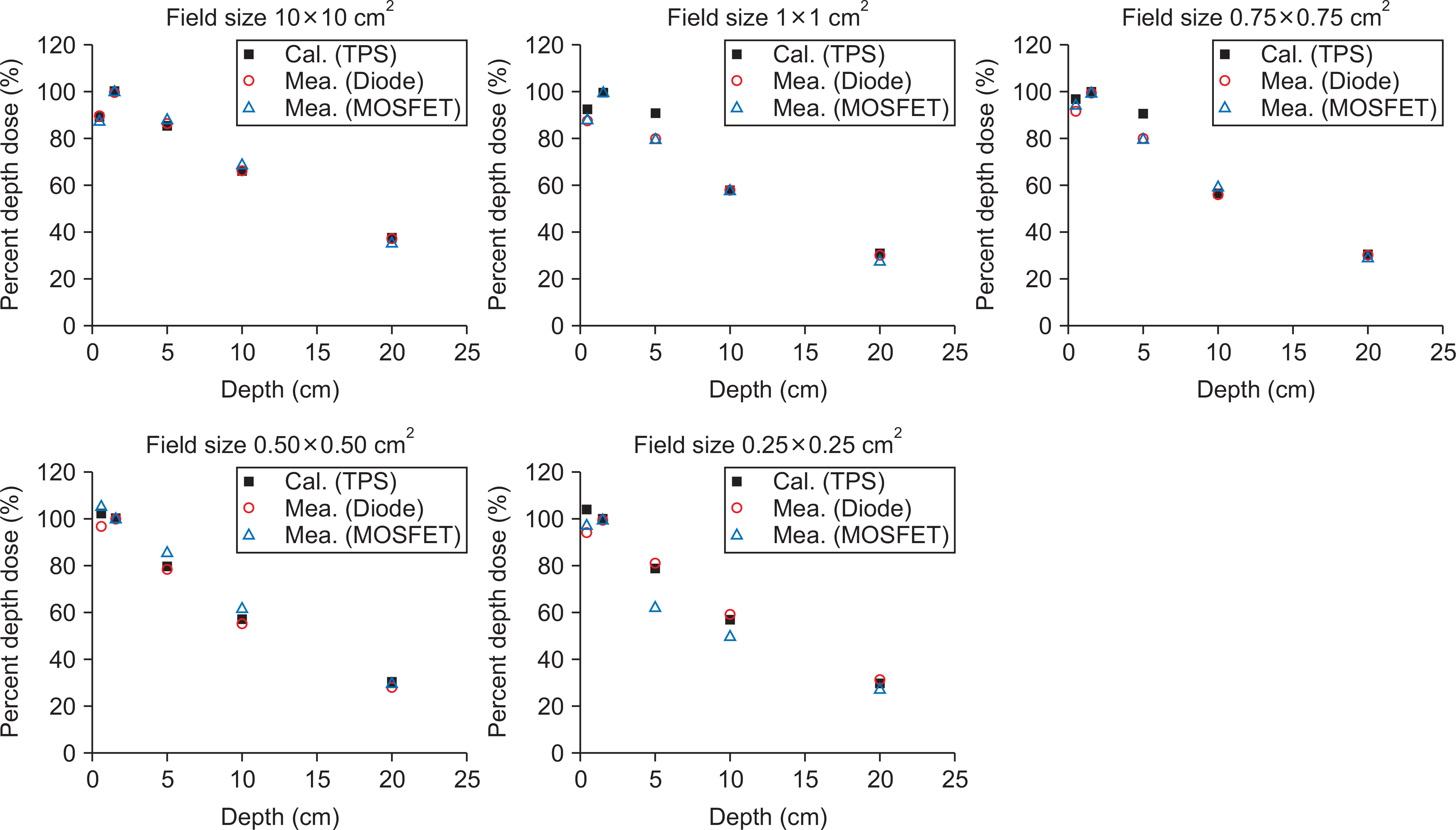
Fig. 1 Calculated and measured percent depth doses (PDDs) at various depths equal to 0.5 cm, 5 cm, 10 cm, 20 cm, and at the depth of the maximum dose (Dmax) for the 6 MV photon beam, and for the field sizes of 10×10 cm2, 1×1 cm2, 0.75×0.75 cm2, 0.50×0.50 cm2, and 0.25×0.25 cm2. The value of Dmax for 6 MV is 1.5 cm. TPS stands for the calculated PDDs using the Eclipse, while Diode and MOSFET stand for measured PDDs using diode and MOSFET detectors, respectively.
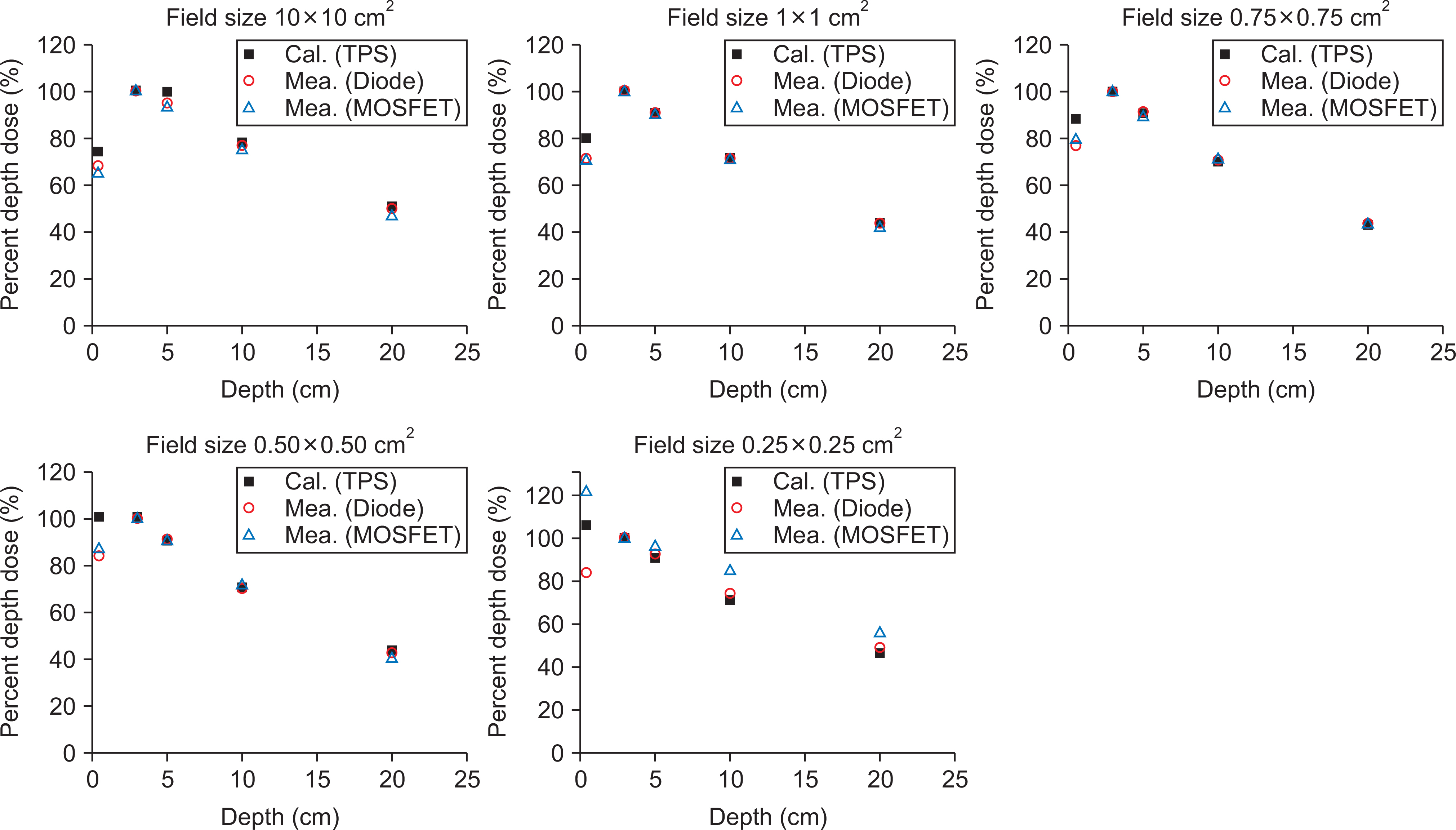
Fig. 2 Calculated and measured percent depth doses (PDDs) at various depths of 0.5 cm, 5 cm, 10 cm, 20 cm, and at the depth of the maximum dose (Dmax) for 15 MV photon beams for the field sizes of 10×10 cm2, 1×1 cm2, 0.75×0.75 cm2, 0.50×0.50 cm2, and 0.25×0.25 cm2. The value of Dmax for 15 MV is 3 cm. TPS stands for the calculated PDDs using Eclipse while Diode and MOSFET stand for measured PDDs using diode and MOSFET detector, respectively.
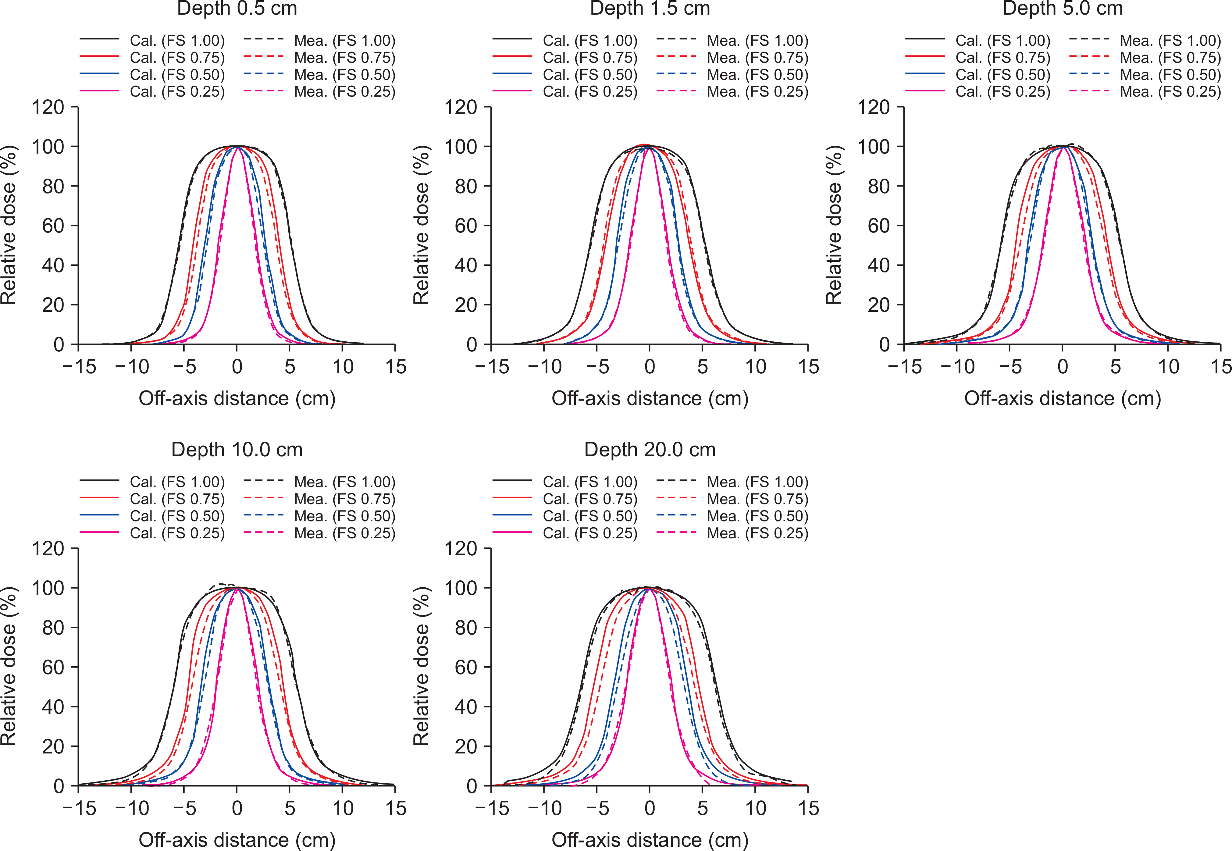
Fig. 3 Calculated and measured lateral profiles at various depths of 0.5 cm, 5 cm, 10 cm, 20 cm, and at the depth of the maximum dose (Dmax) for the 6 MV photon beam for field sizes of 1×1 cm2, 0.75×0.75 cm2, 0.50×0.50 cm2, and 0.25×0.25 cm2. The value of Dmax for 6 MV is 1.5 cm. The calculated and measured lateral profiles are plotted using solid and dashed lines, respectively.
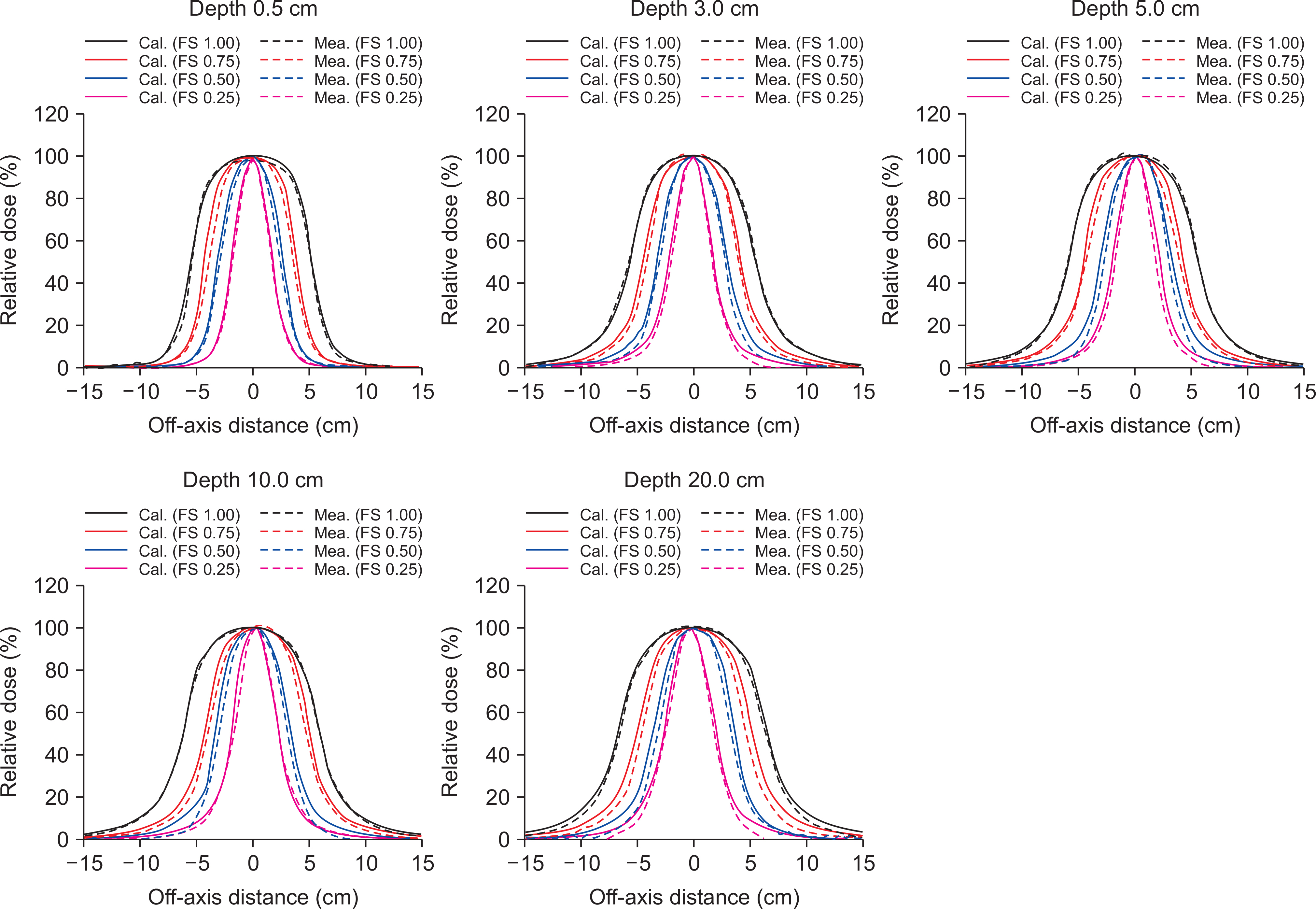
Fig. 4 Calculated and measured lateral profiles at various depths of 0.5 cm, 5 cm, 10 cm, 20 cm, and at a depth of maximum dose (Dmax) for the 6 MV photon beam for the field sizes of 1×1 cm2, 0.75×0.75 cm2, 0.50×0.50 cm2, and 0.25×0.25 cm2. The value of Dmax for 15 MV is 3 cm. The calculated and measured lateral profiles are plotted using solid and dashed lines, respectively.
Reference
-
1.Ezzell GA., Galvin JM., Low D, et al. Guidance document on delivery, treatment planning, and clinical implementation of IMRT: report of the IMRT Subcommittee of the AAPM Radiation Therapy Committee. Med phys. 2003. 30(8):2089–115.
Article2.Zhang P., Happersett L., Hunt M., Jackson A., Zelefsky M., Mageras G. Volumetric modulated arc therapy: planning and evaluation for prostate cancer cases. Int J Radiat Oncol Biol Phys. 2010. 76(5):1456–62.
Article3.Evans JD., Hansen CC., Tollefson MK., Hallemeier CL. Stereotactic body radiation therapy for medically inoperable, clinically localized, urothelial carcinoma of the renal pelvis: A case report. Adv Radia Oncol. 2018. 3(1):57–61.
Article4.Alagar AG., Mani GK., Karunakaran K. Percentage depth dose calculation accuracy of model based algorithms in high energy photon small fields through heterogeneous media and comparison with plastic scintillator dosimetry. J Appl Clin Med Phys. 2016. 17(1):132–42.
Article5.Fogliata A., Cozzi L. Dose calculation algorithm accuracy for small fields in non-homogeneous media: The lung SBRT case. Phys Med. 2017. 44:157–62.
Article6.Adamczyk M., Fundowicz M. Stereotactic body radiation therapy for liver metastasis—case report and review of the literature. The role of patient preparation, treatment planning and its delivery. J Cancer Res Ther. 2014. 10(3):519–25.7.Godson HF., Ravikumar M., Sathiyan S., Ganesh KM., Pon-malar YR., Varatharaj C. Analysis of small field percent depth dose and profiles: Comparison of measurements with various detectors and effects of detector orientation with different jaw settings. J Med Phys. 2016. 41(1):12–20.8.Reggiori G., Mancosu P., Suchowerska N, et al. Characterization of a new unshielded diode for small field dosimetry under flattening filter free beams. Phys Med. 2016. 32(2):408–13.
Article9.Lechner W., Palmans H., Solkner L., Grochowska P., Georg D. Detector comparison for small field output factor measurements in flattening filter free photon beams. Radiotherapy and oncology: Radiother Oncol. 2013. 109(3):356–60.
Article10.Cranmer-Sargison G., Weston S., Evans JA., Sidhu NP., Thwaites DI. Implementing a newly proposed Monte Carlo based small field dosimetry formalism for a comprehensive set of diode detectors. Med phys. 2011. 38(12):6592–602.
Article11.Charles PH., Crowe SB., Kairn T, et al. Monte Carlo-based diode design for correction-less small field dosimetry. Phys Med Biol. 2013. 58(13):4501–12.
Article12.Amin MN., Heaton R., Norrlinger B., Islam MK. Small field electron beam dosimetry using MOSFET detector. J Appl Clin Med Phys. 2010. 12(1):3267.
Article13.Gonzalez-Lopez A., Vera-Sanchez JA., Lago-Martin JD. Small fields measurements with radiochromic films. J Med Phys. 2015. 40(2):61–7.
Article14.Morales JE., Hill R., Crowe SB., Kairn T., Trapp JV. A comparison of surface doses for very small field size x-ray beams: Monte Carlo calculations and radiochromic film measurements. Australas Phys Eng Sci Med. 2014. 37(2):303–9.
Article15.Ralston A., Liu P., Warrener K., Mckenzie D., Suchowerska N. Small field diode correction factors derived using an air core fibre optic scintillation dosimeter and EBT2 film. Phys Med Biol. 2012. 57(9):2587–602.
Article16.Scott AJ., Nahum AE., Fenwick JD. Using a Monte Carlo model to predict dosimetric properties of small radiotherapy photon fields. Med phys. 2008. 35(10):4671–84.
Article17.Scott AJ., Nahum AE., Fenwick JD. Monte Carlo modeling of small photon fields: quantifying the impact of focal spot size on source occlusion and output factors, and exploring miniphantom design for small-field measurements. Med phys. 2009. 36(7):3132–44.
Article18.Francescon P., Kilby W., Satariano N., Cora S. Monte Carlo simulated correction factors for machine specific reference field dose calibration and output factor measurement using fixed and iris collimators on the CyberKnife system. Phys Med Biol. 2012. 57(12):3741–58.
Article19.Pantelis E., Moutsatsos A., Zourari K, et al. On the output factor measurements of the CyberKnife iris collimator small fields: Experimental determination of the k(Q(clin),Q(msr)) (f(clin),f(msr)) correction factors for microchamber and diode detectors. Med phys. 2012. 39(8):4875–85.20.Ciancaglioni I., Marinelli M., Milani E, et al. Dosimetric characterization of a synthetic single crystal diamond detector in clinical radiation therapy small photon beams. Med phys. 2012. 39(7):4493–501.
Article21.Laub WU., Crilly R. Clinical radiation therapy measurements with a new commercial synthetic single crystal diamond detector. J Appl Clin Med Phys. 2014. 15(6):4890.
Article22.Massillon JLG., Cueva-Procel D., Diaz-Aguirre P., Rodriguez-Ponce M., Herrera-Martinez F. Dosimetry for small fields in stereotactic radiosurgery using gafchromic MD-V2-55 film, TLD-100 and alanine dosimeters. PloS one. 2013. 8(5):e63418.23.Crop F., Reynaert N., Pittomvils G, et al. The influence of small field sizes, penumbra, spot size and measurement depth on perturbation factors for microionization chambers. Phys Med Biol. 2009. 54(9):2951–69.
Article24.Parwaie W., Refahi S., Ardekani MA., Farhood B. Different Dosimeters/Detectors Used in Small-Field Dosimetry: Pros and Cons. J Med Signals Sens. 2018. 8(3):195–203.25.Novotny J Jr.., Bhatnagar JP., Quader MA., Bednarz G., Lun-sford LD., Huq MS. Measurement of relative output factors for the 8 and 4 mm collimators of Leksell Gamma Knife Perfexion by film dosimetry. Med phys. 2009. 36(5):1768–74.
Article26.Qin Y., Gardner SJ., Kim J, et al. Technical Note: Evaluation of plastic scintillator detector for small field stereotactic patient-specific quality assurance. Med phys. 2017. 44(10):5509–16.
Article
- Full Text Links
-
- Actions
-
Cited
- CITED
-
- Close
- Share
-
- Similar articles
-
- Dosimetric Verifications of the Output Factors in the Small Field Less Than 3 cm2 Using the Gafchromic EBT2 Films and the Various Detectors
- Study on the Small Fields Dosimetry for High Energy Photon-based Radiation Therapy
- Dosimetric Characteristics of Edge Detector(TM) in Small Beam Dosimetry
- Treatment Planning and Dosimetry of Small Radiation Fields for Stereotactic Radiosurgery
- Dose Characteristics of Small Radiation Fields for 6MV X-ray of Linear Accelerator
