Intraoperative Monitoring of Hypoglossal Nerve Using Hypoglossal Motor Evoked Potential in Infratentorial Tumor Surgery: A Report of Two Cases
- Affiliations
-
- 1Department of Rehabilitation Medicine, Seoul National University Hospital, Seoul, Korea. hangil_seo@snuh.org
- 2Department of Neurosurgery, Seoul National University Hospital, Seoul, Korea.
- KMID: 2432207
- DOI: http://doi.org/10.5535/arm.2018.42.2.352
Abstract
- The hypoglossal nerve (CN XII) may be placed at risk during posterior fossa surgeries. The use of intraoperative monitoring (IOM), including the utilization of spontaneous and triggered electromyography (EMG), from tongue muscles innervated by CN XII has been used to reduce these risks. However, there were few reports regarding the intraoperative transcranial motor evoked potential (MEP) of hypoglossal nerve from the tongue muscles. For this reason, we report here two cases of intraoperative hypoglossal MEP monitoring in brain surgery as an indicator of hypoglossal deficits. Although the amplitude of the MEP was reduced in both patients, only in the case 1 whose MEP was disappeared demonstrated the neurological deficits of the hypoglossal nerve. Therefore, the disappearance of the hypoglossal MEP recorded from the tongue, could be considered a predictor of the postoperative hypoglossal nerve deficits.
MeSH Terms
Figure
-
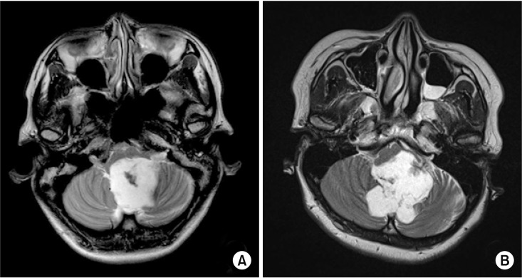
Fig. 1 The preoperative T2-weighted images of brain magnetic resonance imaging showed 3.4 cm×3.6 cm×5.7 cm mass in case 1 (A) and 5.2 cm×5.4 cm×4.8 cm mass in case 2 (B) at the fourth ventricle.
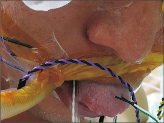
Fig. 2 The recording needle electrodes for motor evoked potential and spontaneous electromyography of the hypoglossal nerve were inserted on the midline of the oral tongue.
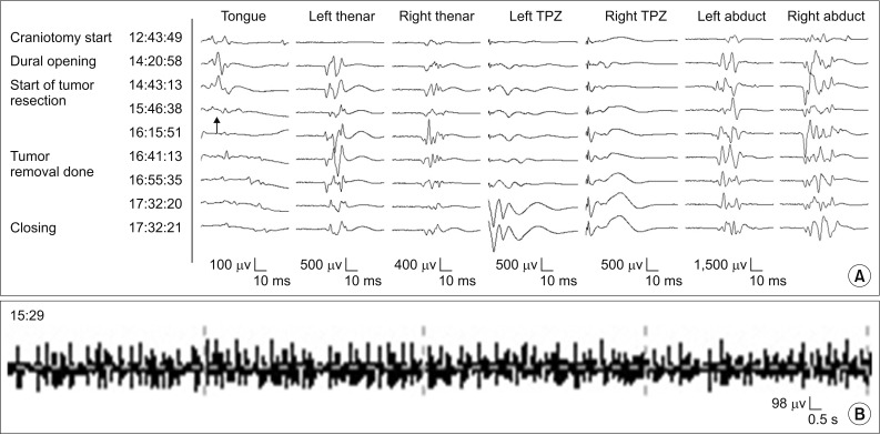
Fig. 3 The tongue and other muscles MEP (A) and spontaneous tongue EMG (B) recorded in case 1. During surgery, the tonic discharge from the spontaneous EMG of tongue muscles was monitored at 15:29 (B) and 20 minutes later, the amplitude of the tongue's MEP was disappeared at 15:46 (arrow) (A). There were no abnormal findings in other muscles. MEP, motor evoked potential; EMG, electromyography.
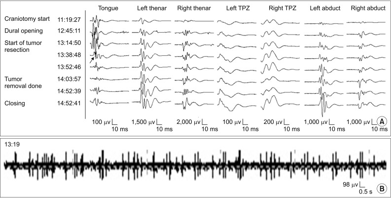
Fig. 4 The tongue and other muscles MEP (A) and spontaneous tongue EMG (B) recorded in case 2. The small tonic discharge was detected from the spontaneous EMG of tongue muscles at 13:19 (B). After 20 minutes, the amplitude of tongue's MEP was reduced at 13:38 to the 50% of its baseline (arrow) (A). There were no abnormal findings in other muscles. MEP, motor evoked potential; EMG, electromyography.
Reference
-
1. Jahangiri FR, Minhas M, Jane J. Preventing lower cranial nerve injuries during fourth ventricle tumor resection by utilizing intraoperative neurophysiological monitoring. Neurodiagn J. 2012; 52:320–332. PMID: 23301282.2. Schlake HP, Goldbrunner RH, Milewski C, Krauss J, Trautner H, Behr R, et al. Intra-operative electromyographic monitoring of the lower cranial motor nerves (LCN IX-XII) in skull base surgery. Clin Neurol Neurosurg. 2001; 103:72–82. PMID: 11516548.
Article3. Topsakal C, Al-Mefty O, Bulsara KR, Williford VS. Intraoperative monitoring of lower cranial nerves in skull base surgery: technical report and review of 123 monitored cases. Neurosurg Rev. 2008; 31:45–53. PMID: 17957398.
Article4. Holdefer RN, Kinney GA, Robinson LR, Slimp JC. Alternative sites for intraoperative monitoring of cranial nerves X and XII during intracranial surgeries. J Clin Neurophysiol. 2013; 30:275–279. PMID: 23733092.
Article5. Ishikawa M, Kusaka G, Takashima K, Kamochi H, Shinoda S. Intraoperative monitoring during surgery for hypoglossal schwannoma. J Clin Neurosci. 2010; 17:1053–1056. PMID: 20488709.
Article6. Kullmann M, Tatagiba M, Liebsch M, Feigl GC. Evaluation of the predictive value of intraoperative changes in motor-evoked potentials of caudal cranial nerves for the postoperative functional outcome. World Neurosurg. 2016; 95:329–334. PMID: 27485529.
Article7. Fukuda M, Takao T, Hiraishi T, Yajima N, Saito A, Fujii Y. Novel devices for intraoperative monitoring of glossopharyngeal and vagus nerves during skull base surgery. Surg Neurol Int. 2013; 4:97. PMID: 23956940.
Article8. Akagami R, Dong CC, Westerberg BD. Localized transcranial electrical motor evoked potentials for monitoring cranial nerves in cranial base surgery. Neurosurgery. 2005; 57(1 Suppl):78–85. PMID: 15987572.
Article
- Full Text Links
-
- Actions
-
Cited
- CITED
-
- Close
- Share
-
- Similar articles
-
- Idiopathic Isolated Hypoglossal Nerve Palsy After Upper Respiratory Infection
- Hypoglossal Neurinoma without Preoperative Hypoglossal Nerve Palsy - Report of 2 Cases -
- Isolated Unilateral Hypoglossal Nerve Palsy Following Transoral Endotracheal Intubation for Endoscopic Sinus Surgery
- Delayed Isolated Hypoglossal Nerve Palsy after Submandibular Gland Surgery
- A Case of Transient Glossopharyngeal and Hypoglossal Nerve Palsy after Laryngomicrosurgery
