Ann Dermatol.
2016 Feb;28(1):115-116. 10.5021/ad.2016.28.1.115.
Increasing Numbers of Mast Cells in Skin Lesions of Hyperpigmented Mycosis Fungoides with Large-Cell Transformation
- Affiliations
-
- 1Department of Dermatology, Tokyo Medical and Dental University, Graduate School of Medicine, Tokyo, Japan. k.igawa.derm@tmd.ac.jp
- KMID: 2429494
- DOI: http://doi.org/10.5021/ad.2016.28.1.115
Abstract
- No abstract available.
MeSH Terms
Figure
-
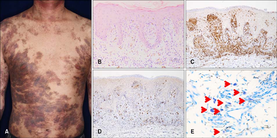
Fig. 1 (A) Clinical manifestation. Hyperpigmented plaques on the trunk. (B) Histology of the lesional skin of a hyperpigmented plaque. Slight acanthosis with atypical lymphocytic infiltration within the epidermis and upper dermis, containing abundant melanophages (H&E, ×100). (C, D) Immunohistochemical analysis (×100). (C) CD4 staining and (D) CD8 staining. (E) Mast cells in lesional skin. Red arrowheads indicate mast cells (toluidine blue, ×400).
Reference
-
1. Yamamoto T, Katayama I, Nishioka K. Role of mast cell and stem cell factor in hyperpigmented mycosis fungoides. Blood. 1997; 90:1338–1340.
Article2. Lee JS, Yun SJ, Lee JB, Kim SJ, Won YH, Lee SC. A case of hyperpigmented mycosis fungoides: a rare variant. J Eur Acad Dermatol Venereol. 2007; 21:983–985.
Article3. Erbil H, Sezer E, Koseoglu D, Filiz N, Kurumlu Z, Bülent Taştan H, et al. Hyperpigmented mycosis fungoides: a case report. J Eur Acad Dermatol Venereol. 2007; 21:982–983.
Article4. Pavlovsky L, Mimouni D, Amitay-Laish I, Feinmesser M, David M, Hodak E. Hyperpigmented mycosis fungoides: an unusual variant of cutaneous T-cell lymphoma with a frequent CD8+ phenotype. J Am Acad Dermatol. 2012; 67:69–75.
Article
- Full Text Links
-
- Actions
-
Cited
- CITED
-
- Close
- Share
-
- Similar articles
-
- A Case of Transformation of Mycosis Fungoides to Large Cell Lymphoma
- A Case of Ki-1 Positive Large-Cell Lymphoma Transformed from Mycosis Fungoides
- A Case of Primary Cutaneous CD30 Positive Anaplastic Large Cell Lymphoma in a Patient with Mycosis Fungoides
- A Case of Fluorine-18-Fluorodeoxyglucose Positron Emission Tomography/Computed Tomography Findings of Mycosis Fungoides with Large Cell Transformation Involving Cutaneous Lesions, and Metastasis to Lymph Node and Viscera
- Immunohistochemical Study of T lymphocytes Infiltrated in Mycosis Fungoides: Comparison with Psoriasis and Eczema
