Atypical Fracture of the Ulna Associated with 3 Years of Bisphosphonate Medication
- Affiliations
-
- 1Department of Orthopedic Surgery, Yeungnam University Medical Center, Daegu, Korea. radiorth@ynu.ac.kr
- KMID: 2429177
- DOI: http://doi.org/10.4055/jkoa.2018.53.3.277
Abstract
- There have been many studies regarding the relationship between long-term use of bisphosphonate and atypical femoral fractures in the literature. However, studies regarding atypical fractures of the upper limbs are severely limited, especially in Korea. Here, we report an atypical fracture of the ulna in a patient with bisphosphonate medication for a relatively short period of 3 years without any history of fractures at other sites.
Keyword
Figure
-
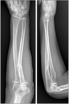
Figure 1 Anteroposterior and lateral radiographs show a minimally displaced fracture of the left proximal ulna. Cortical thickening and marginal sclerosis are observed at the transverse fracture line.

Figure 2 Sagittal image of computed tomography also shows cortical thickening and marginal sclerosis at the transverse fracture line.

Figure 3 Whole body bone scan shows focal hot uptake in the left proximal ulna (arrows) and right femur shaft (arrows). Intravenous injection of the bone scan agent (technetium-99m methoxy diphosphonate) was performed at the right forearm (arrowheads).
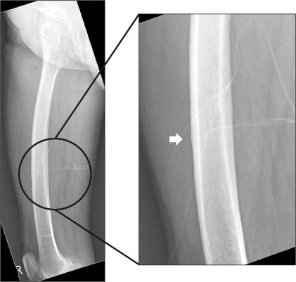
Figure 4 Lateral radiograph of the right femur shows diffuse cortical thickening and incomplete fracture in the right femoral shaft (arrow).
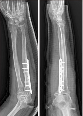
Figure 5 Anteroposterior and lateral radiographs of the left forearm taken postoperatively.

Figure 6 (A) Anteroposterior and lateral radiographs of the left forearm taken 1 year postoperatively show bone union of the ulna fracture. (B) Lateral radiograph of the right femur taken 1 year after the initial forearm injury show no definite change (arrow).
Reference
-
1. Odvina CV, Zerwekh JE, Rao DS, Maalouf N, Gottschalk FA, Pak CY. Severely suppressed bone turnover: a potential complication of alendronate therapy. J Clin Endocrinol Metab. 2005; 90:1294–1301.
Article2. Neviaser AS, Lane JM, Lenart BA, Edobor-Osula F, Lorich DG. Low-energy femoral shaft fractures associated with alendronate use. J Orthop Trauma. 2008; 22:346–350.
Article3. Giusti A, Hamdy NA, Papapoulos SE. Atypical fractures of the femur and bisphosphonate therapy: a systematic review of case/case series studies. Bone. 2010; 47:169–180.4. Shane E, Burr D, Abrahamsen B, et al. Atypical subtrochanteric and diaphyseal femoral fractures: second report of a task force of the American Society for Bone and Mineral Research. J Bone Miner Res. 2014; 29:1–23.
Article5. Saita Y, Ishijima M, Mogami A, et al. The incidence of and risk factors for developing atypical femoral fractures in Japan. J Bone Miner Metab. 2015; 33:311–318.
Article6. Schilcher J, Aspenberg P. Incidence of stress fractures of the femoral shaft in women treated with bisphosphonate. Acta Orthop. 2009; 80:413–415.
Article7. Tan SH, Saseendar S, Tan BH, Pawaskar A, Kumar VP. Ulnar fractures with bisphosphonate therapy: a systematic review of published case reports. Osteoporos Int. 2015; 26:421–429.
Article8. Chiang GS, Koh KW, Chong TW, Tan BY. Stress fracture of the ulna associated with bisphosphonate therapy and use of walking aid. Osteoporos Int. 2014; 25:2151–2154.
Article9. Tang ZH, Kumar VP. Alendronate-associated ulnar and tibial fractures: a case report. J Orthop Surg (Hong Kong). 2011; 19:370–372.
Article10. Stathopoulos KD, Kosmidis C, Lyritis GP. Atypical fractures of the femur and ulna and complications of fracture healing in a 76-year-old woman with Sjögren's syndrome. J Musculoskelet Neuronal Interact. 2011; 11:208–211. quiz 211.
- Full Text Links
-
- Actions
-
Cited
- CITED
-
- Close
- Share
-
- Similar articles
-
- Atypical Fracture of the Proximal Shaft of the Ulna Associated with Prolonged Bisphosphonate Therapy
- Treatment of Atypical Ulnar Fracture Associated with Bisphosphonate Therapy - A Case Report -
- Ulnar Insufficiency Fractures in Patients on Prolonged Bisphosphonate Therapy: A Case Report
- Atypical femoral neck fracture after prolonged bisphosphonate therapy
- Management Challenges in Atypical Femoral Fractures: A Case Report
