Ann Dermatol.
2018 Dec;30(6):744-746. 10.5021/ad.2018.30.6.744.
A Case of Indeterminate Dendritic Cell Tumor: A Rare Neoplasm with Langerhans Cell Lineage
- Affiliations
-
- 1Department of Dermatology, Seoul National University College of Medicine, Seoul, Korea. khcho@snu.ac.kr
- KMID: 2428941
- DOI: http://doi.org/10.5021/ad.2018.30.6.744
Abstract
- No abstract available.
MeSH Terms
Figure
-
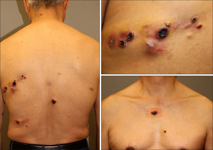
Fig. 1 Multiple reddish brown necrotic and crusted nodules on the back and chest.
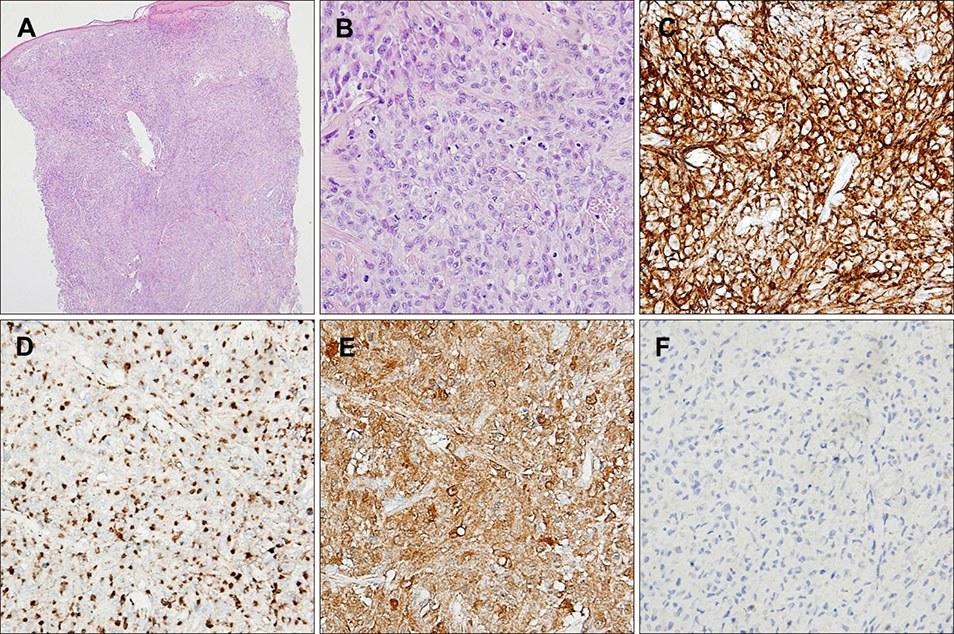
Fig. 2 (A) Aggregates of tumor cells resembling Langerhans cells throughout dermis (H&E, ×40). (B) Tumors cells exhibiting varied shaped nuclei and abundant cytoplasm (H&E, ×400). (C) Positive immunohistochemical staining for CD1a (×400), (D) CD68 (×400), and (E) S100 proteins (×400). (F) Negative immunohistochemical staining for langerin (×400).
Reference
-
1. Rezk SA, Spagnolo DV, Brynes RK, Weiss LM. Indeterminate cell tumor: a rare dendritic neoplasm. Am J Surg Pathol. 2008; 32:1868–1876.2. Mo X, Guo W, Ye H. Primary indeterminate dendritic cell tumor of skin correlated to mosquito bite. Medicine (Baltimore). 2015; 94:e1443.
Article3. Roh J, Kim SW, Park CS. Indeterminate dendritic cell tumor: a case report of a rare langerhans cell lineage disease. J Pathol Transl Med. 2016; 50:78–81.
Article4. Cheuk W, Cheung FY, Lee KC, Chan JK. Cutaneous indeterminate dendritic cell tumor with a protracted relapsing clinical course. Am J Surg Pathol. 2009; 33:1261–1263.
Article5. Contreras F, Fonseca E, Gamallo C, Burgos E. Multiple self-healing indeterminate cell lesions of the skin in an adult. Am J Dermatopathol. 1990; 12:396–401.
Article
- Full Text Links
-
- Actions
-
Cited
- CITED
-
- Close
- Share
-
- Similar articles
-
- Recurrent Indeterminate Dendritic Cell Tumor of the Skin
- Indeterminate Dendritic Cell Tumor: A Case Report of a Rare Langerhans Cell Lineage Disease
- A Case of Indeterminate Cell Histiocytosis in an Infant
- Macrophage/dendritic Cell Marker Staining Characteristics of Langerhans cell Granulomatosis(Histiocytosis X)
- A Case of Solitary Indeterminate Cell Histiocytoma
