Indeterminate Dendritic Cell Tumor: A Case Report of a Rare Langerhans Cell Lineage Disease
- Affiliations
-
- 1Department of Pathology, Asan Medical Center, University of Ulsan College of Medicine, Seoul, Korea. csikpark@amc.seoul.kr
- KMID: 2211411
- DOI: http://doi.org/10.4132/jptm.2015.07.03
Abstract
- No abstract available.
MeSH Terms
Figure
-
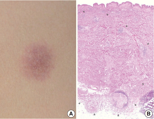
Fig. 1. (A) An ill-defined, 1.2-cm, round, erythematous subcutaneous nodule on the left flank. (B) The ill-defined tumor is mainly located in the dermis and subcutis. The margin of the tumor is marked by arrowheads.

Fig. 2. (A) There is patchy infiltration by aggregates of the tumor cells. Tumor cells are usually monotonous and have indistinct cell borders. Clusters of lymphocytes are admixed with tumor cells in focal areas without eosinophils. (B) The constituent cells have ovoid cell morphology with abundant eosinophilic cytoplasm. (C) Their nuclei are oval and sometimes indented. Nuclear grooves are frequently seen. Although occasional enlarged nuclei are identified, mitoses are rarely seen. (D) There is no epidermal involvement.
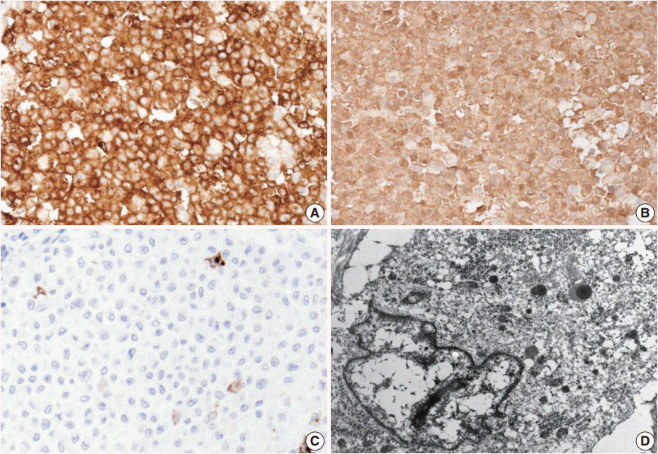
Fig. 3. The neoplastic cells diffusely express CD1a (A) and S100 protein (B). (C) Langerin (CD207) is typically negative. (D) No Birbeck granules are seen on transmission electron microscopy (×8,000).
Cited by 2 articles
-
A Case of Indeterminate Dendritic Cell Tumor: A Rare Neoplasm with Langerhans Cell Lineage
Jungyoon Moon, Ji Hoon Yang, Jaewon Lee, Jong Seo Park, Kwang Hyun Cho
Ann Dermatol. 2018;30(6):744-746. doi: 10.5021/ad.2018.30.6.744.Recurrent Indeterminate Dendritic Cell Tumor of the Skin
Jin Woo Joo, Taek Chung, Yoon Ah Cho, Sang Kyum Kim
J Pathol Transl Med. 2018;52(4):243-247. doi: 10.4132/jptm.2018.03.27.
Reference
-
1. Weiss LM, Chan JK, Fletcher CD. Other rare dendritic cell tumours. In : Swerdlow SH, Campo E, Harris NL, editors. WHO classification of tumours of haematopoietic and lymphoid tissues. 4th ed. Lyon: IARC Press;2008. p. 365.2. Ghanadan A, Kamyab K, Ramezani M, et al. Indeterminate cell histiocytosis: report of a case. Acta Med Iran. 2014; 52:788–90.3. Rezk SA, Spagnolo DV, Brynes RK, Weiss LM. Indeterminate cell tumor: a rare dendritic neoplasm. Am J Surg Pathol. 2008; 32:1868–76.4. Vener C, Soligo D, Berti E, et al. Indeterminate cell histiocytosis in association with later occurrence of acute myeloblastic leukaemia. Br J Dermatol. 2007; 156:1357–61.
Article5. Wang CH, Chen GS. Indeterminate cell histiocytosis: a case report. Kaohsiung J Med Sci. 2004; 20:24–30.
Article6. Calonje E, Brenn T, Lazar A, McKee PH. McKee’s pathology of the skin with clinical correlations. 4th ed. Edinburgh: Saunders;2011. p. 1398.7. Bakry OA, Samaka RM, Kandil MA, Younes SF. Indeterminate cell histiocytosis with naive cells. Rare Tumors. 2013; 5:e13.8. Bohn OL, Ruiz-Argüelles G, Navarro L, Saldivar J, Sanchez-Sosa S. Cutaneous Langerhans cell sarcoma: a case report and review of the literature. Int J Hematol. 2007; 85:116–20.
Article9. Chikwava K, Jaffe R. Langerin (CD207) staining in normal pediatric tissues, reactive lymph nodes, and childhood histiocytic disorders. Pediatr Dev Pathol. 2004; 7:607–14.10. Lau SK, Chu PG, Weiss LM. Immunohistochemical expression of Langerin in Langerhans cell histiocytosis and non-Langerhans cell histiocytic disorders. Am J Surg Pathol. 2008; 32:615–9.
Article
- Full Text Links
-
- Actions
-
Cited
- CITED
-
- Close
- Share
-
- Similar articles
-
- Recurrent Indeterminate Dendritic Cell Tumor of the Skin
- A Case of Indeterminate Dendritic Cell Tumor: A Rare Neoplasm with Langerhans Cell Lineage
- A Case of Indeterminate Cell Histiocytosis in an Infant
- A Case of Solitary Indeterminate Cell Histiocytoma
- Macrophage/dendritic Cell Marker Staining Characteristics of Langerhans cell Granulomatosis(Histiocytosis X)
