Yonsei Med J.
2005 Dec;46(6):806-811.
Comparison of the Predictive Value of Myelography, Computed Tomography and MRI on the Treadmill Test in Lumbar Spinal Stenosis
- Affiliations
-
- 1Department of Orthop Surg, Yongdong Severance Hospital, Yonsei University College of Medicine, Seoul, Korea.
- 2Department of Orthop Surg, Cha University College of Medicine, Sungnam, Korea.
- 3Department of Orthop Surg, National Health Insurance Corporation Ilsan Hospital, Koyang, Korea.
- 4Department of Orthop Surg, Jaesaeng Hospital, Sungnam, Korea.
- 5The BK 21 Project for Medical Science in Yonsei University, Seoul, Korea.
Abstract
- To date, there have been no prospective, objective studies comparing the accuracy of the MRI, myelo-CT and myelography. The purpose of this study is to compare the diagnostic and predictive values of MRIs, myelo-CTs, and myelographies. Myelographies with dynamic motion views, myelo-CTs, MRIs and exercise treadmill tests were performed in 35 cases. The narrowest AP diameter of the dural sac was measured by myelography. At the pathologic level, dural cross-sectional area (D-CSA) was calculated in the MRI and Myelo-CT. The time to the first symptoms (TAF) and the total ambulation time (TAT) were measured during the exercise treadmill test and used as the standard in the comparison of correlation between radiographic parameters and walking capacity. The mean D-CSA by CT was 58.3 mm2 and 47.6 mm2 by MRI. All radiographic parameters such as AP diameters and D-CSA have no correlation to TAF or TAT (p > 0.05). Our data showed no statistically significant differences in the correlation of the patients' walking capacity to the severity of stenosis as assessed by myelography, myelo-CT and MRI.
Keyword
MeSH Terms
Figure
-
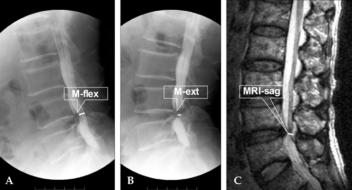
Fig. 1 Lateral projections of functional myelogram from a patient with lumbar spinal stenosis in (A) provoked flexion, and (B) extended position. Note the increase of the sagittal diameter at the third and fourth level in the flexed position.
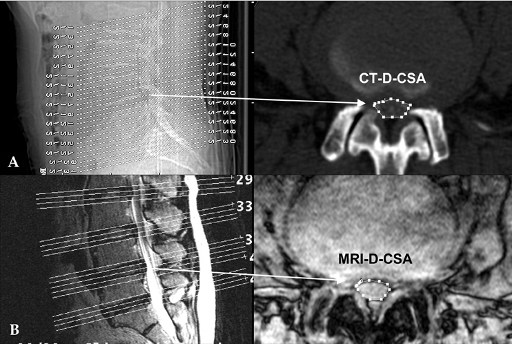
Fig. 2 (A) The CT scan slices chosen were parallel to the lower vertebral endplate. The pathologic D-CSA was measured at the disc level. (B) The MRI scan slices chosen were parallel to the lower vertebral endplate. The pathologic D-CSA was measured at the disc level.
Reference
-
1. Herno A, Airaksinen O, Saari T, Miettinen H. The predictive value of preoperative myelography in lumbar spinal stenosis. Spine. 1994. 19:1335–1338.2. Sortland O, Magnaes B, Hauge T. Functional myelography with metrizamide in the diagnosis of lumbar spinal stenosis. Acta Radiol. 1977. 355:Suppl. 42–54.3. Bolender NF, Schonstrom NS, Spengler DM. Role of computed tomography and myelography in the diagnosis of central spinal stenosis. J Bone Joint Surg Am. 1985. 67:240–246.4. Bischoff RJ, Rodriguez RP, Gupta K, Righi A, Dalton JE, Whitecloud TS. A comparison of computed tomography-myelography, magnetic resonance imaging and myelography in the diagnosis of herniated nucleus pulposus and spinal stnosis. J Spinal Dis. 1993. 6:289–295.5. Schonstrom N, Willen J. Imaging lumbar spinal stenosis. Radiol Clin North Am. 2001. 39:31–53.6. Herzog RJ. Lumbar spine: Spinal stenosis. I.C.L. #152. 2002. In : American Academy of Orthopaedic Surgeon Annual Meeting;7. Spivak JM. Current concepts review: degenerative lumbar spinal stenosis. J Bone Joint Surg Am. 1998. 80:1053–1066.8. Willen J, Danielson B, Gaulitz A, Niklason T, Schonstrom N, Hansson T. Dynamic effects on the lumbar spinal canal: axially loaded CT-myelography and MRI in patients with sciatica and/or neurogenic claudication. Spine. 1997. 22:2968–2976.9. Deen HG, Zimmerman RS, Lyons MK, McPhee MC, Verheijde JL, Lemens SM. Use of the exercise treadmill to measure baseline functional status and surgical outcome in patients with severe lumbar spinal stenosis. Spine. 1998. 23:244–248.10. Deen HG, Zimmerman RS, Lyons MK, McPhee MC, Verheijde JL, Lemens SM. Measurement of exercise tolerance on the treadmill in patients with symptomatic lumbar spinal stenosis: a useful indicator of functional status and surgical outcome. J Neurosurg. 1995. 83:27–30.11. Walters BC. Bean JR, editor. Clinical practice parameter development in neurosurgery. Neurosurgery in transition: the socioeconomic transformation of neurological surgery. 1998. Baltimore: Williams & Wilkins;99–111.12. Burr BA, Resnick D, Syklawer R, Haghighi P. Fluid-fluid levels in a unicameral bone cyst: CT and MR findings. J Comput Assist Tomogr. 1993. 17:134–136.13. Turner JA, Ersek M, Herron L, Deyo R. Surgery for lumbar spinal stenosis: attempted meta-analysis of the literature. Spine. 1992. 17:1–8.14. Lee HM, Kim NH, Kim HJ, Chung IH. Morphometric study of the lumbar spinal canal in the Korean population. Spine. 1995. 20:1679–1684.
- Full Text Links
-
- Actions
-
Cited
- CITED
-
- Close
- Share
-
- Similar articles
-
- Comparison of Magnetic Resonance Imaging and Computed Tomography-Myelography for Quantitative Evaluation of Lumbar Intracanalar Cross-Section
- The Pathomorphologic Study of Spinal Stenosis as Seen on CT - Myelography of the Lumbar
- Diagnosis of Herniated Lumbar Disk with Computed Tomography
- Interobserver Reliability between MRI, CT-myelogram and Myelogram in the Evaluation of Lumbar Spinal Stenosis
- Diagnostic Significance of Myelo-Enhanced Computerized Tomography(MECT) in Spinal Stenosis
