Comparison of clinical and histological characteristics of orthodontic tooth movement into recent and healed extraction sites combined with corticotomy in rats
- Affiliations
-
- 1Orthodontic Section, Department of Preventive Dentistry, Faculty of Dentistry, Prince of Songkla University, Hat Yai, Songkhla, Thailand. samruaj@hotmail.com
- 2Dental Department Maharat Nakhon Ratchasima Hospital, Nakhon Ratchasima, Thailand.
- KMID: 2427778
- DOI: http://doi.org/10.4041/kjod.2018.48.6.405
Abstract
OBJECTIVE
This study was performed to investigate the rate of tooth movement and histological characteristics of extraction sockets those were subjected to corticotomy.
METHODS
A split-mouth randomized controlled trial experiment was designed. Thirty-two adult, male Wistar rats were divided into 2 groups: healing extraction socket (H) and recent extraction socket (R); these groups were randomly classified into 4 subgroups (0/7/21/60 days). The first maxillary molar was extracted on 1 side and 2 months were allowed for complete bone healing; then, the corresponding molar was extracted on the other side and surgical intervention was performed at the mid-alveolar point of the first maxillary molar. Ten grams of continuous force was applied. The outcomes measured were rate of tooth movement, percentage of periodontal space and histological evaluation. The rate of tooth movement was calculated as the measured distance divided by the duration of molar movement. Histomorphometric evaluations were performed on the second and third maxillary molars. The Wilcoxon signed rank test was used to compare differences between the two groups.
RESULTS
There were no significant differences in the rates of tooth movement between H and R groups at any of the 4 time points. The histological appearance and percentage of periodontal space between the R and H groups also demonstrated no significant differences.
CONCLUSIONS
The rates of orthodontic tooth movement into recent and healed socket sites did not differ between the groups. Histological analysis of tooth movement revealed regional acceleration during every time period.
Keyword
Figure
-
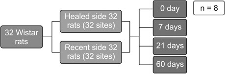
Figure 1 Diagram of the study design.
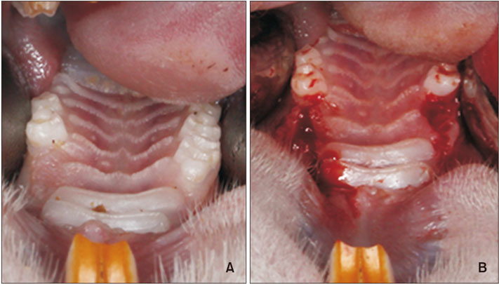
Figure 2 Clinical photographs. A, Complete healing of the healed side at 2 months after extraction. B, After extraction at the recent side and decortication on both sides.
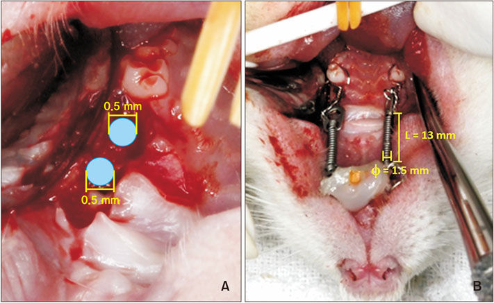
Figure 3 Decortication and orthodontic protocol. A, Midalveolar two-point decortication (diameter, 0.5 mm) at mesial to second maxillary molar. B, Closed coil spring (length, 8 mm; diameter, 1.5 mm) insertion to mesialize the maxillary second molar.
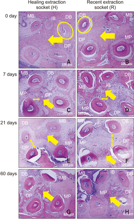
Figure 4 Representative images of histological appearance at the level of the apical third of the maxillary second molar. A, C, E, and G, Healing extraction socket at days 0, 7, 21, and 60, respectively. B, D, F, and H, Recent extraction socket at days 0, 7, 21, and 60, respectively. A and B (Day 0), Full-filled interradicular bone (arrow), continuous and well-defined periodontal space (circle). C and D (Day 7), Non-mineralized tissue and periodontal space (single-ended arrow) was expanded, while the interradicular bone (double-ended arrow) was reduced. E and F (Day 21), Nearly interradicular bone (single-ended arrow) was replaced with non-mineralized tissue, continuous with the periodontal space (double-ended arrow). G and H (Day 60), Non-mineralized tissue was partially replaced with rebuilt interradicular bone (single-ended arrow), while the periodontal space was reduced. MB, Mesiobuccal root; IB, intermediate buccal root; DB, distobuccal root; DP, distopalatal root; MP, mesiopalatal root. Hematoxylin and eosin stain.
Reference
-
1. Kole H. Surgical operations on the alveolar ridge to correct occlusal abnormalities. Oral Surg Oral Med Oral Pathol. 1959; 12:515–529. concl.
Article2. Frost HM. The biology of fracture healing. An overview for clinicians Part II. Clin Orthop Relat Res. 1989; (248):294–309.3. Frost HM. The biology of fracture healing. An overview for clinicians Part I. Clin Orthop Relat Res. 1989; (248):283–293.4. Yaffe A, Fine N, Binderman I. Regional accelerated phenomenon in the mandible following mucoperiosteal flap surgery. J Periodontol. 1994; 65:79–83.
Article5. Sebaoun JD, Kantarci A, Turner JW, Carvalho RS, Van Dyke TE, Ferguson DJ. Modeling of trabecular bone and lamina dura following selective alveolar decortication in rats. J Periodontol. 2008; 79:1679–1688.
Article6. Wang L, Lee W, Lei DL, Liu YP, Yamashita DD, Yen SL. Tisssue responses in corticotomy- and osteotomy-assisted tooth movements in rats: histology and immunostaining. Am J Orthod Dentofacial Orthop. 2009; 136:770.e1–770.e11. discussion 770-1.
Article7. Mostafa YA, Mohamed Salah Fayed M, Mehanni S, ElBokle NN, Heider AM. Comparison of corticotomy-facilitated vs standard tooth-movement techniques in dogs with miniscrews as anchor units. Am J Orthod Dentofacial Orthop. 2009; 136:570–577.
Article8. Iino S, Sakoda S, Ito G, Nishimori T, Ikeda T, Miyawaki S. Acceleration of orthodontic tooth movement by alveolar corticotomy in the dog. Am J Orthod Dentofacial Orthop. 2007; 131:448.e1–448.e8.
Article9. Liou EJ, Huang CS. Rapid canine retraction through distraction of the periodontal ligament. Am J Orthod Dentofacial Orthop. 1998; 114:372–382.
Article10. Häsler R, Schmid G, Ingervall B, Gebauer U. A clinical comparison of the rate of maxillary canine retraction into healed and recent extraction sites--a pilot study. Eur J Orthod. 1997; 19:711–719.
Article11. Diedrich P, Wehrbein H. Orthodontic retraction into recent and healed extraction sites. A histologic study. J Orofac Orthop. 1997; 58:90–99.12. Murphey WH Jr. Oxytetracycline microfluorescent comparison of orthodontic retraction into recent and healed extraction sites. Am J Orthod. 1970; 58:215–239.
Article13. Hsieh YD, Devlin H, Roberts C. Early alveolar ridge osteogenesis following tooth extraction in the rat. Arch Oral Biol. 1994; 39:425–428.
Article14. Astrand P, Carlsson GE. Changes in the alveolar process after extractions in the white rat. A histologic and fluorescence microscopic study. Acta Odontol Scand. 1969; 27:113–127.
Article15. Kim SJ, Park YG, Kang SG. Effects of corticision on paradental remodeling in orthodontic treatment. Angle Orthod. 2009; 79:284–291.16. Reitan K, Kvam E. Comparative behavior of human and animal tissue during experimental tooth movement. Angle Orthod. 1971; 41:1–14.17. Rygh P. Hyalinization of the periodontal ligament incident to orthodontic tooth movement. Nor Tannlaegeforen Tid. 1974; 84:352–357.
Article
- Full Text Links
-
- Actions
-
Cited
- CITED
-
- Close
- Share
-
- Similar articles
-
- Corticotomy for orthodontic tooth movement
- Mode of tooth movement according to the timing of orthodontic force application after extraction
- Corticotomy and the Intrusive Tooth Movement
- The effectiveness of corticotomy and piezocision on canine retraction: A systematic review
- Use of corticotomy for canine and molar retraction
