Evaluation of changes in the maxillary alveolar bone after incisor intrusion
- Affiliations
-
- 1Department of Orthodontics, Faculty of Dentistry, Hacettepe University, Ankara, Turkey. ezgibaytorun@hotmail.com
- KMID: 2427774
- DOI: http://doi.org/10.4041/kjod.2018.48.6.367
Abstract
OBJECTIVE
This study was performed to investigate the changes in alveolar bone after maxillary incisor intrusion and to determine the related factors in deep-bite patients.
METHODS
Fifty maxillary central incisors of 25 patients were evaluated retrospectively. The maxillary incisors in Group I (12 patients; mean age, 16.51 ± 1.32 years) were intruded with a base-arch, while those in Group II (13 patients; mean age, 17.47 ± 2.71 years) were intruded with miniscrews. Changes in the alveolar envelope were assessed using pre-intrusion and post-intrusion cone-beam computed tomography images. Labial, palatal, and total bone thicknesses were evaluated at the crestal (3 mm), midroot (6 mm), and apical (9 mm) levels. Buccal and palatal alveolar crestal height, buccal bone height, and the prevalence of dehiscence were evaluated. Two-way repeated measure ANOVA was used to determine the significance of the changes. Pearson's correlation coefficient analysis was performed to assess the relationship between dental and alveolar bone measurement changes.
RESULTS
Upper incisor inclination and intrusion changes were significantly greater in Group II than in Group I. With treatment, the alveolar bone thickness at the labial bone thickness (LBT, 3 and 6 mm) decreased significantly in Group II (p < 0.001) as compared to Group I. The LBT change at 3 mm was strongly and positively correlated with the amount of upper incisor intrusion (r = 0.539; p = 0.005).
CONCLUSIONS
Change in the labial inclination and the amount of intrusion should be considered during upper incisor intrusion, as these factors increase the risk of alveolar bone loss.
MeSH Terms
Figure
-

Figure 1 Location of alveolar bone thickness measurements. LBT, Labial bone thickness; TBT, total bone thicknesses; PBT, palatal bone thickness.
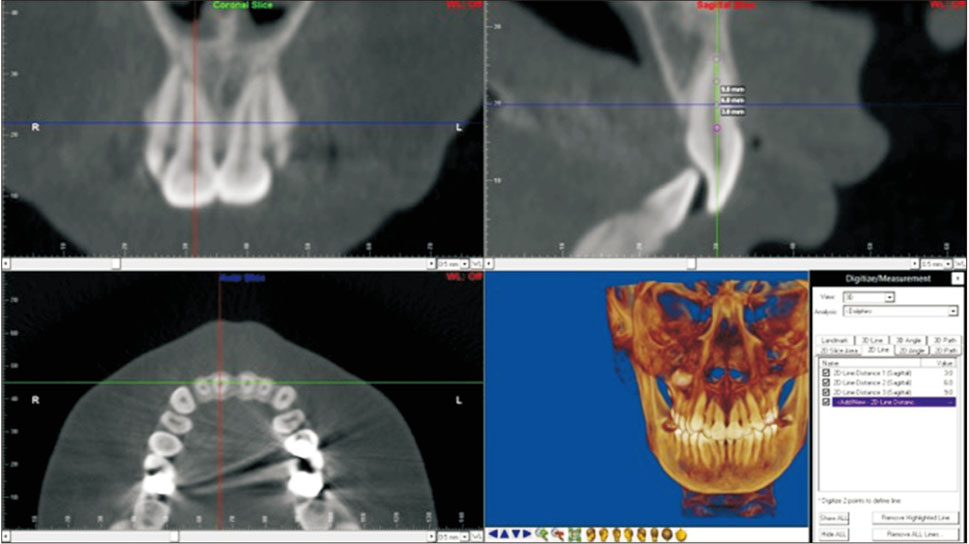
Figure 2 Orientation of all 3 planes of space on cone-beam computed tomography.

Figure 3 Location of alveolar bone height measurements. PACH, Palatal alveolar crestal height; BACH, buccal alveolar crestal height; BBH, buccal bone height.
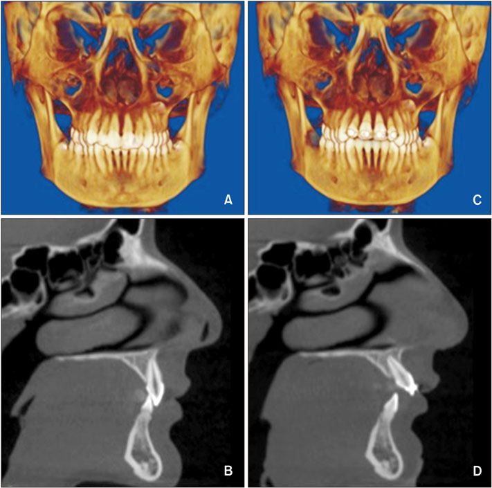
Figure 4 Cone-beam computed tomography images of a representative case. A, B, Pretreatment. C, D, Post-treatment.
Cited by 2 articles
-
Cone-beam computed tomographic evaluation of mandibular incisor alveolar bone changes for the intrusion arch technique: A retrospective cohort research
Lin Lu, Jiaping Si, Zhikang Wang, Xiaoyan Chen
Korean J Orthod. 2024;54(2):79-88. doi: 10.4041/kjod23.173.Combined anterior and posterior miniscrews increase apical root resorption of maxillary incisors in protrusion and premolar extraction cases
Zhizun Wang, Li Mei, Zhenxing Tang, Dong Wu, Yue Zhou, Ehab A. Abdulghani, Yuan Li, Wei Zheng, Yu Li
Korean J Orthod. 2025;55(1):26-36. doi: 10.4041/kjod24.136.
Reference
-
1. Nauert K, Berg R. Evaluation of labio-lingual bony support of lower incisors in orthodontically untreated adults with the help of computed tomography. J Orofac Orthop. 1999; 60:321–334.
Article2. Verna C, Zaffe D, Siciliani G. Histomorphometric study of bone reactions during orthodontic tooth movement in rats. Bone. 1999; 24:371–379.
Article3. Garlock DT, Buschang PH, Araujo EA, Behrents RG, Kim KB. Evaluation of marginal alveolar bone in the anterior mandible with pretreatment and posttreatment computed tomography in nonextraction patients. Am J Orthod Dentofacial Orthop. 2016; 149:192–201.
Article4. Melsen B. Biological reaction of alveolar bone to orthodontic tooth movement. Angle Orthod. 1999; 69:151–158.5. Nanda R, Kuhlberg A. Management of deep overbite malocclusion. In : Nanda R, editor. Biomechanics and esthetic strategies in clinical orthodontics. 1st ed. St. Louis: Elsevier Saunders;2005. p. 131–155.6. Burzin J, Nanda R. The stability of deep overbite correction. In : Nanda R, Burstone CJ, editors. Retention and stability in orthodontics. Philadelphia: WB Saunders;1993.7. Burstone CJ. Biomechanics of deep overbite correction. Semin Orthod. 2001; 7:26–33.
Article8. Steiner GG, Pearson JK, Ainamo J. Changes of the marginal periodontium as a result of labial tooth movement in monkeys. J Periodontol. 1981; 52:314–320.
Article9. Batenhorst KF, Bowers GM, Williams JE Jr. Tissue changes resulting from facial tipping and extrusion of incisors in monkeys. J Periodontol. 1974; 45:660–668.
Article10. Thongudomporn U, Charoemratrote C, Jearapongpakorn S. Changes of anterior maxillary alveolar bone thickness following incisor proclination and extrusion. Angle Orthod. 2015; 85:549–554.
Article11. Kaied IB, Tanielian RH. Comparative radiographic evaluation of the alveolar bone support changes after incisal intrusion. Orthodontics (Chic.). 2012; 13:60–71.12. Timock AM, Cook V, McDonald T, Leo MC, Crowe J, Benninger BL, et al. Accuracy and reliability of buccal bone height and thickness measurements from cone-beam computed tomography imaging. Am J Orthod Dentofacial Orthop. 2011; 140:734–744.
Article13. Handelman CS. The anterior alveolus: its importance in limiting orthodontic treatment and its influence on the occurrence of iatrogenic sequelae. Angle Orthod. 1996; 66:95–109. discussion 109-10.14. Vardimon AD, Oren E, Ben-Bassat Y. Cortical bone remodeling/tooth movement ratio during maxillary incisor retraction with tip versus torque movements. Am J Orthod Dentofacial Orthop. 1998; 114:520–529.
Article15. Ludlow JB, Laster WS, See M, Bailey LJ, Hershey HG. Accuracy of measurements of mandibular anatomy in cone beam computed tomography images. Oral Surg Oral Med Oral Pathol Oral Radiol Endod. 2007; 103:534–542.
Article16. Kim Y, Park JU, Kook YA. Alveolar bone loss around incisors in surgical skeletal Class III patients. Angle Orthod. 2009; 79:676–682.
Article17. Lee KM, Kim YI, Park SB, Son WS. Alveolar bone loss around lower incisors during surgical orthodontic treatment in mandibular prognathism. Angle Orthod. 2012; 82:637–644.
Article18. Karagöz A. Derin örtülü kapanışlı olgularda üst keser intrüzyonunun konik ışınlı bilgisayarlı tomografi ile incelenmesi. Ankara, Turkey: Hacettepe University;2013.19. Sarikaya S, Haydar B, Ciğer S, Ariyürek M. Changes in alveolar bone thickness due to retraction of anterior teeth. Am J Orthod Dentofacial Orthop. 2002; 122:15–26.
Article20. Persson RE, Hollender LG, Laurell L, Persson GR. Horizontal alveolar bone loss and vertical bone defects in an adult patient population. J Periodontol. 1998; 69:348–356.
Article21. Mischkowski RA, Pulsfort R, Ritter L, Neugebauer J, Brochhagen HG, Keeve E, et al. Geometric accuracy of a newly developed cone-beam device for maxillofacial imaging. Oral Surg Oral Med Oral Pathol Oral Radiol Endod. 2007; 104:551–559.
Article22. Tian YL, Liu F, Sun HJ, Lv P, Cao YM, Yu M, et al. Alveolar bone thickness around maxillary central incisors of different inclination assessed with cone-beam computed tomography. Korean J Orthod. 2015; 45:245–252.
Article23. Yodthong N, Charoemratrote C, Leethanakul C. Factors related to alveolar bone thickness during upper incisor retraction. Angle Orthod. 2013; 83:394–401.
Article24. Ahn HW, Moon SC, Baek SH. Morphometric evaluation of changes in the alveolar bone and roots of the maxillary anterior teeth before and after en masse retraction using cone-beam computed tomography. Angle Orthod. 2013; 83:212–221.
Article25. Picanço PR, Valarelli FP, Cançado RH, de Freitas KM, Picanço GV. Comparison of the changes of alveolar bone thickness in maxillary incisor area in extraction and non-extraction cases: computerized tomography evaluation. Dental Press J Orthod. 2013; 18:91–98.
Article26. Fuhrmann R. Three-dimensional evaluation of periodontal remodeling during orthodontic treatment. Semin Orthod. 2002; 8:23–28.
Article27. Cobo J, Argüelles J, Puente M, Vijande M. Dentoalveolar stress from bodily tooth movement at different levels of bone loss. Am J Orthod Dentofacial Orthop. 1996; 110:256–262.
Article28. Bimstein E, Crevoisier RA, King DL. Changes in the morphology of the buccal alveolar bone of protruded mandibular permanent incisors secondary to orthodontic alignment. Am J Orthod Dentofacial Orthop. 1990; 97:427–430.
Article29. Kalha A. Gingival recession and labial movement of lower incisors. Evid Based Dent. 2013; 14:21–22.
Article30. Aziz T, Flores-Mir C. A systematic review of the association between appliance-induced labial movement of mandibular incisors and gingival recession. Aust Orthod J. 2011; 27:33–39.31. Cho SM, Choi SH, Sung SJ, Yu HS, Hwang CJ. The effects of alveolar bone loss and miniscrew position on initial tooth displacement during intrusion of the maxillary anterior teeth: Finite element analysis. Korean J Orthod. 2016; 46:310–322.
Article32. Sun Z, Smith T, Kortam S, Kim DG, Tee BC, Fields H. Effect of bone thickness on alveolar bone-height measurements from cone-beam computed tomography images. Am J Orthod Dentofacial Orthop. 2011; 139:e117–e127.
Article
- Full Text Links
-
- Actions
-
Cited
- CITED
-
- Close
- Share
-
- Similar articles
-
- Cone-beam computed tomographic evaluation of mandibular incisor alveolar bone changes for the intrusion arch technique: A retrospective cohort research
- The effects of alveolar bone loss and miniscrew position on initial tooth displacement during intrusion of the maxillary anterior teeth: Finite element analysis
- Three-dimensional finite element analysis on intrusion of upper anterior teeth by three-piece base arch appliance according to alveolar bone loss
- Three-dimensional evaluation of maxillary anterior alveolar bone for optimal placement of miniscrew implants
- Reconstruction of the alveolar cleft with gingivo-vestibular-mucoperiosteal flap
