The effects of alveolar bone loss and miniscrew position on initial tooth displacement during intrusion of the maxillary anterior teeth: Finite element analysis
- Affiliations
-
- 1Private Practice, Namyangju, Korea.
- 2Department of Orthodontics, The Institute of Craniofacial Deformity, College of Dentistry, Yonsei University, Seoul, Korea. hwang@yuhs.ac
- 3Division of Orthodontics, Department of Dentistry, Asan Medical Center, University of Ulsan College of Medicine, Seoul, Korea.
- KMID: 2352293
- DOI: http://doi.org/10.4041/kjod.2016.46.5.310
Abstract
OBJECTIVE
The aim of this study was to determine the optimal loading conditions for pure intrusion of the six maxillary anterior teeth with miniscrews according to alveolar bone loss.
METHODS
A three-dimensional finite element model was created for a segment of the six anterior teeth, and the positions of the miniscrews and hooks were varied after setting the alveolar bone loss to 0, 2, or 4 mm. Under 100 g of intrusive force, initial displacement of the individual teeth in three directions and the degree of labial tilting were measured.
RESULTS
The degree of labial tilting increased with reduced alveolar bone height under the same load. When a miniscrew was inserted between the two central incisors, the amounts of medial-lateral and anterior-posterior displacement of the central incisor were significantly greater than in the other conditions. When the miniscrews were inserted distally to the canines and an intrusion force was applied distal to the lateral incisors, the degree of labial tilting and the amounts of displacement of the six anterior teeth were the lowest, and the maximum von Mises stress was distributed evenly across all the teeth, regardless of the bone loss.
CONCLUSIONS
Initial tooth displacement similar to pure intrusion of the six maxillary anterior teeth was induced when miniscrews were inserted distal to the maxillary canines and an intrusion force was applied distal to the lateral incisors. In this condition, the maximum von Mises stresses were relatively evenly distributed across all the teeth, regardless of the bone loss.
Keyword
Figure
-

Figure 1 Three-dimensional finite element models and the coordination system. A, Model of the maxillary teeth. B, C, and D, Lateral views of the models with 0, 2, and 4 mm of alveolar bone loss respectively. A +x value was defined as the lateral direction, −y as the posterior direction, and +z as the superior direction, to analyze the displacement of individual teeth.

Figure 2 Force vectors for the intrusion of the six-tooth anterior segment (green box). Black and white circles, miniscrew heads; red arrows, load vectors.
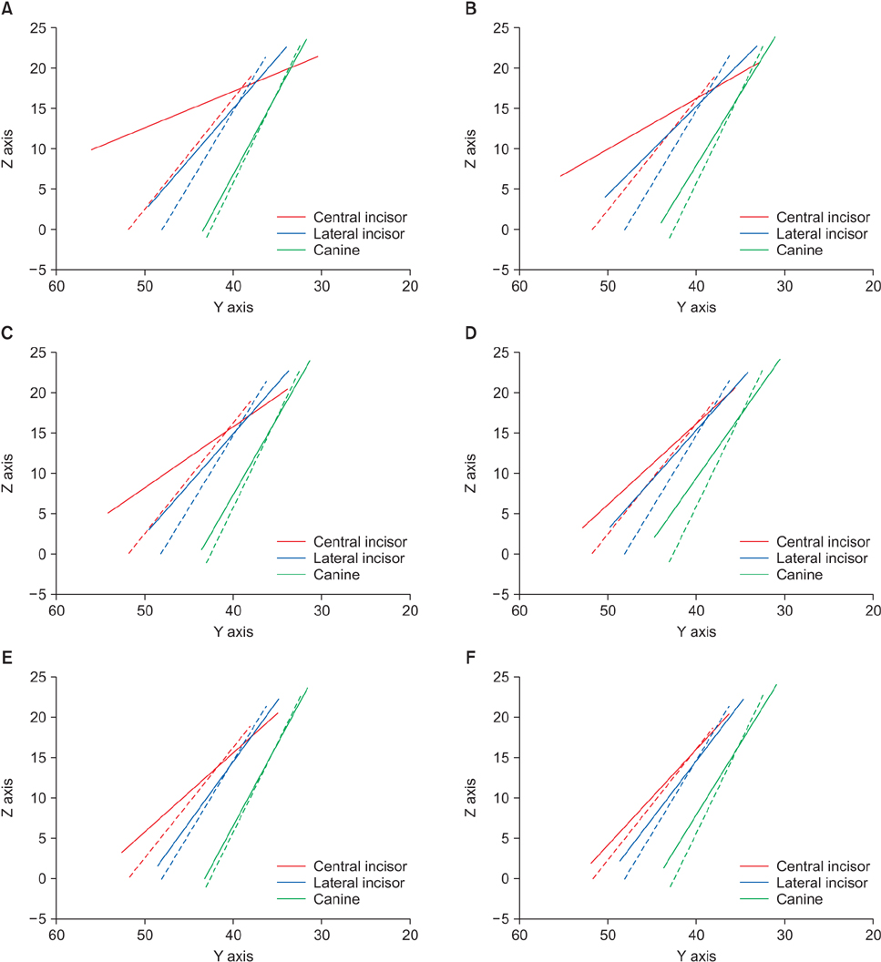
Figure 3 Sagittal view of the displacement of the anterior teeth without bone loss in the six-tooth anterior segment. Each line represents the longitudinal axis of each tooth. The displacement is scaled up 1,000-fold to enhance visibility. Each figure refers to the each loading condition (See also Figure 2). Dotted line, before the displacement of the individual teeth; solid line, after the displacement of the individual teeth.

Figure 4 Sagittal view of the displacement of the anterior teeth with 2 mm of bone loss in the six-tooth anterior segment. Each line represents the longitudinal axis of each tooth. The displacement is scaled up 1,000-fold to enhance visibility. Each figure refers to the each loading condition (See also Figure 2). Dotted line, before the displacement of the individual teeth; solid line, after the displacement of the individual teeth.
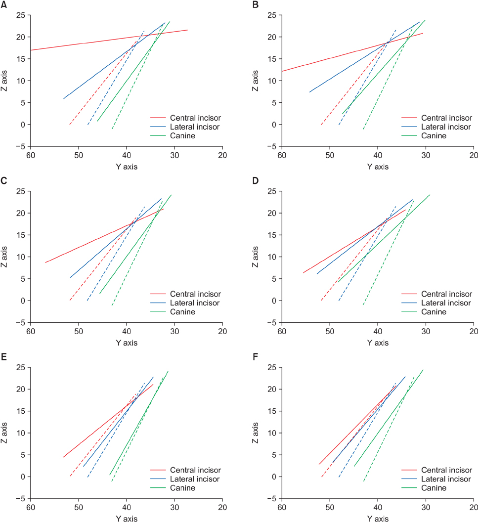
Figure 5 Sagittal view of the displacement of the anterior teeth with 4 mm of bone loss in the six-tooth anterior segment. Each line represents the longitudinal axis of each tooth. The displacement is scaled up 1,000-fold to enhance visibility. Each figure refers to the each loading condition (See also Figure 2). Dotted line, before the displacement of the individual teeth; solid line, after the displacement of the individual teeth.

Figure 6 Comparison of the maximum compressive stress distributions in periodontal ligaments with 4 mm of bone loss. Blue color indicates high-stress distribution, and red color indicates low-stress distribution. Each figure refers to the each loading condition (See also Figure 2).
Cited by 3 articles
-
Finite-element analysis of the center of resistance of the mandibular dentition
A-Ra Jo, Sung-Seo Mo, Kee-Joon Lee, Sang-Jin Sung, Youn-Sic Chun
Korean J Orthod. 2017;47(1):21-30. doi: 10.4041/kjod.2017.47.1.21.Prediction of optimal bending angles of a running loop to achieve bodily protraction of a molar using the finite element method
Woon-Kuk Ryu, Jae Hyun Park, Kiyoshi Tai, Yukio Kojima, Youngjoo Lee, Jong-Moon Chae
Korean J Orthod. 2018;48(1):3-10. doi: 10.4041/kjod.2018.48.1.3.Evaluation of changes in the maxillary alveolar bone after incisor intrusion
Ezgi Atik, Hande Gorucu-Coskuner, Bengisu Akarsu-Guven, Tulin Taner
Korean J Orthod. 2018;48(6):367-376. doi: 10.4041/kjod.2018.48.6.367.
Reference
-
1. Kang DY, Choi SH, Jung YS, Hwang CJ. Interdisciplinary treatment for an adult patient with anterior open bite, severe periodontitis, and intellectual disability. J Craniofac Surg. 2015; 26:e240–e244.
Article2. Serio FG, Hawley CE. Periodontal trauma and mobility. Diagnosis and treatment planning. Dent Clin North Am. 1999; 43:37–44.3. Rohatgi S, Narula SC, Sharma RK, Tewari S, Bansal P. A study on clinical attachment loss and gingival inflammation as etiologic factors in pathologic tooth migration. Niger J Clin Pract. 2011; 14:449–453.
Article4. Towfighi PP, Brunsvold MA, Storey AT, Arnold RM, Willman DE, McMahan CA. Pathologic migration of anterior teeth in patients with moderate to severe periodontitis. J Periodontol. 1997; 68:967–972.
Article5. Kanomi R. Mini-implant for orthodontic anchorage. J Clin Orthod. 1997; 31:763–767.6. Nishimura M, Sannohe M, Nagasaka H, Igarashi K, Sugawara J. Nonextraction treatment with temporary skeletal anchorage devices to correct a Class II Division 2 malocclusion with excessive gingival display. Am J Orthod Dentofacial Orthop. 2014; 145:85–94.
Article7. Choi JH, Yu HS, Lee KJ, Park YC. Three-dimensional evaluation of maxillary anterior alveolar bone for optimal placement of miniscrew implants. Korean J Orthod. 2014; 44:54–61.
Article8. Lee KJ, Park YC, Hwang CJ, Kim YJ, Choi TH, Yoo HM, et al. Displacement pattern of the maxillary arch depending on miniscrew position in sliding mechanics. Am J Orthod Dentofacial Orthop. 2011; 140:224–232.
Article9. Cobo J, Argüelles J, Puente M, Vijande M. Dentoalveolar stress from bodily tooth movement at different levels of bone loss. Am J Orthod Dentofacial Orthop. 1996; 110:256–262.
Article10. Cobo J, Sicilia A, Argüelles J, Suárez D, Vijande M. Initial stress induced in periodontal tissue with diverse degrees of bone loss by an orthodontic force: tridimensional analysis by means of the finite element method. Am J Orthod Dentofacial Orthop. 1993; 104:448–454.
Article11. Cattaneo PM, Dalstra M, Melsen B. The finite element method: a tool to study orthodontic tooth movement. J Dent Res. 2005; 84:428–433.
Article12. Coolidge ED. The thickness of the human periodontal membrane. J Am Dent Assoc. 1937; 24:1260–1270.
Article13. Kronfeld R. Histologic study of the influence of function on the human periodontal membrane. J Am Dent Assoc. 1931; 18:1242–1274.
Article14. Park HK, Sung EH, Cho YS, Mo SS, Chun YS, Lee KJ. 3-D FEA on the intrusion of mandibular anterior segment using orthodontic miniscrews. Korean J Orthod. 2011; 41:384–398.
Article15. Sung EH, Kim SJ, Chun YS, Park YC, Yu HS, Lee KJ. Distalization pattern of whole maxillary dentition according to force application points. Korean J Orthod. 2015; 45:20–28.
Article16. Sung SJ, Baik HS, Moon YS, Yu HS, Cho YS. A comparative evaluation of different compensating curves in the lingual and labial techniques using 3D FEM. Am J Orthod Dentofacial Orthop. 2003; 123:441–450.
Article17. Vollmer D, Bourauel C, Maier K, Jäger A. Determination of the centre of resistance in an upper human canine and idealized tooth model. Eur J Orthod. 1999; 21:633–648.
Article18. Reimann S, Keilig L, Jäger A, Bourauel C. Biomechanical finite-element investigation of the position of the centre of resistance of the upper incisors. Eur J Orthod. 2007; 29:219–224.
Article19. Lee EH, Yu HS, Lee KJ, Park YC. Three dimensional finite element analysis of continuous and segmented arches with use of orthodontic miniscrews. Korean J Orthod. 2011; 41:237–254.
Article20. Mo SS, Kim SH, Sung SJ, Chung KR, Chun YS, Kook YA, et al. Torque control during lingual anterior retraction without posterior appliances. Korean J Orthod. 2013; 43:3–14.
Article21. Burstone CR. Deep overbite correction by intrusion. Am J Orthod. 1977; 72:1–22.
Article22. Ricketts RM. Bioprogressive therapy as an answer to orthodontic needs. Part I. Am J Orthod. 1976; 70:241–268.
Article23. Smith RJ, Burstone CJ. Mechanics of tooth movement. Am J Orthod. 1984; 85:294–307.
Article24. Polat-Ozsoy O, Arman-Ozcirpici A, Veziroglu F. Miniscrews for upper incisor intrusion. Eur J Orthod. 2009; 31:412–416.
Article25. Sia S, Koga Y, Yoshida N. Determining the center of resistance of maxillary anterior teeth subjected to retraction forces in sliding mechanics. An in vivo study. Angle Orthod. 2007; 77:999–1003.
Article26. Sung SJ, Kim IT, Kook YA, Chun YS, Kim SH, Mo SS. Finite-element analysis of the shift in center of resistance of the maxillary dentition in relation to alveolar bone loss. Korean J Orthod. 2009; 39:278–288.
Article27. Dermaut LR, De Munck A. Apical root resorption of upper incisors caused by intrusive tooth movement: a radiographic study. Am J Orthod Dentofacial Orthop. 1986; 90:321–326.
Article28. Parker RJ, Harris EF. Directions of orthodontic tooth movements associated with external apical root resorption of the maxillary central incisor. Am J Orthod Dentofacial Orthop. 1998; 114:677–683.
Article
- Full Text Links
-
- Actions
-
Cited
- CITED
-
- Close
- Share
-
- Similar articles
-
- Three-dimensional finite element analysis on intrusion of upper anterior teeth by three-piece base arch appliance according to alveolar bone loss
- 3-D FEA on the intrusion of mandibular anterior segment using orthodontic miniscrews
- A three-dimensional finite element analysis on the location of center of resistance during intrusion of upper anterior teeth
- Finite-element investigation of the center of resistance of the maxillary dentition
- Influence of changing various parameters in miniscrew-assisted rapid palatal expansion: A three-dimensional finite element analysis
