Alveolar Squamous Cell Metaplasia: Preneoplastic Lesion?
- Affiliations
-
- 1Service d'Anatomie Pathologique, APHP GHU Avicenne, Bobigny, France. adriana.handra-luca@aphp.fr
- 2Universite Paris Nord Sorbonne Cite, Bobigny, France.
- KMID: 2427516
- DOI: http://doi.org/10.4132/jptm.2018.09.07
Abstract
- No abstract available.
MeSH Terms
Figure
-
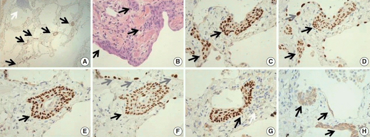
Fig. 1. (A) The lung parenchyma shows a zone of multiple (approximatively 10) p63-positive squamous cell metaplasia (SCM) foci (black arrows for SCM foci, white arrow for lymphocytic focus). (B) On the hematoxylin and eosin stained slide, the lesions consist in a multilayered epithelium composed of basal cuboidal cells, suprabasal cells and superficial spindle-appearing cells (black arrows). (C) The basal and suprabasal cells are immunoreactive for p63 while superficial cells are negative (black arrows for p63+ cells). (D) Thyroid transcription factor 1 is expressed by the cells throughout the entire thickness of the lesion, in both p63+ and p63– cells (black arrows for SCM foci, gray arrow for atypical pneumocyte nuclei). (E, F) A cystic cellular bud (reminiscent of thyroid solid cell nests) is detected in an alveolar septum (black arrows for the SCM bud, gray arrows for binucleated pneumocytes). (G) One of the SCM foci develop at close contact to the lymphocytic infiltrate (p63 immunohistochemistry, black arrow for the SCM focus, white arrow for the lymphcytic infiltrate). To note would be the presence of a binucleation with immunoreactivity to p63 in the SCM focus. (H) Cytokeratin 5/6 is expressed in spindle-appearing cells lining the alveoli and in the SCM foci (black arrows for cytokeratin 5/6+ cells).
Reference
-
1. Song DH, Choi IH, Ha SY, et al. Usual interstitial pneumonia with lung cancer: clinicopathological analysis of 43 cases. Korean J Pathol. 2014; 48:10–6.
Article2. Meyer EC, Liebow AA. Relationship of interstitial pneumonia honeycombing and atypical epithelial proliferation to cancer of the lung. Cancer. 1965; 18:322–51.
Article3. Rosai J. Rosai and Ackerman’s surgical pathology. 10th ed. Philadelphia: Elsevier Mosby;2011.4. Chilosi M, Poletti V, Murer B, et al. Abnormal re-epithelialization and lung remodeling in idiopathic pulmonary fibrosis: the role of deltaN-p63. Lab Invest. 2002; 82:1335–45.5. Zuo W, Zhang T, Wu DZ, et al. p63(+)Krt5(+) distal airway stem cells are essential for lung regeneration. Nature. 2015; 517:616–20.
Article6. Kato E, Takayanagi N, Takaku Y, et al. Incidence and predictive factors of lung cancer in patients with idiopathic pulmonary fibrosis. ERJ Open Res. 2018; 4:00111–2016.
Article7. Krimsky W, Muganlinskaya N, Sarkar S, et al. The changing anatomic position of squamous cell carcinoma of the lung: a new conundrum. J Community Hosp Intern Med Perspect. 2016; 6:33299.
- Full Text Links
-
- Actions
-
Cited
- CITED
-
- Close
- Share
-
- Similar articles
-
- Squamous Metaplasia of the Pleura
- Squamous Metaplasia of the Pleura
- An Image Analytical Study on the Structural Spectrum of Intestinal Metaplasia-Dysplasia-Carcinoma of the Stomach
- Autoradiographic Labeling Index of Tritiated Thymidine in Human Lung Cancer
- Oncogene expressions detected by in situ hybridization of squamous metaplasia, dysplasia and primary lung cancer in human
