J Korean Ophthalmol Soc.
2018 Nov;59(11):1087-1090. 10.3341/jkos.2018.59.11.1087.
Trochlear Nerve Palsy Caused by Quadrigeminal Cistern Lipoma
- Affiliations
-
- 1Department of Ophthalmology, Yeungnam University College of Medicine, Daegu, Korea. mmk@med.yu.ac.kr
- KMID: 2426372
- DOI: http://doi.org/10.3341/jkos.2018.59.11.1087
Abstract
- PURPOSE
To report a case of trochlear nerve palsy caused by quadrigeminal cistern lipoma located in the dorsal midbrain.
CASE SUMMARY
A 65-year-old male visited our clinic for intermittent vertical diplopia over 2-year period. Symptoms of diplopia had worsened over the past two weeks. He had no previous medical history except having had diabetes for 1 month. The best-corrected visual acuity was 20/25 in the right eye and 20/20 in the left eye. Pupillary examination was not remarkable. Extraocular examination showed 4 prism diopters (PD) left hypertropia at distant gaze and 4 PD exotropia at near gaze, with adduction elevation of the left eye. The Bielschowsky head tilt test revealed 6 PD left hypertropia on the left gaze and orthotropia on the right tilt. Fundus examination showed excyclotorsion of the right eye and incyclotorsion of the left eye. Brain magnetic resonance imaging revealed quadrigeminal cistern lipoma. Prism glasses were prescribed to alleviate diplopia, and we followed up the lesions without further treatment.
CONCLUSIONS
Trochlear nerve palsy can be caused by quadrigeminal cistern lipoma; however, it is uncommon for this condition to be caused by a compressive lesion. Prompt neuroimaging can be helpful to rule out the causes of this condition in patients with atypical symptoms.
Keyword
MeSH Terms
Figure
-
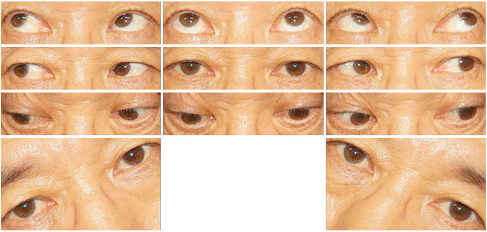
Figure 1 Image of the patient in nine diagnostic position and both side tilting. The patient showed left hypertropia at primary position with elevation in adduction of the left eye. The hypertropia of the left eye increased in left side tilting position.

Figure 2 T1-weighted magnetic resonance imaging (MRI) of brain. (A) Sagittal T1-weighted magnetic resonance imaging (MRI) of brain showed a well-defined hyperintensity suggesting lipoma in median region of quadrigeminal cistern adjacent to the anterior medullary vellum (arrow). (B) Axial T1-weighted MRI image demonstrated lipoma located adjacent to the emerging site of trochlear nerve fiber (arrowhead).
Reference
-
1. Miller NR, Subramanian PS, Patel VR. Walsh and Hoyt's Clinical Neuro-Ophthalmology: the essentials. 3rd ed. Philadelphia: Wolters Kluwer;2016. p. 355–361.2. Kline LB, Foroozan R. Neuro-Ophthalmology Review manual. 7th ed. Thorofare: SLACK;2013. p. 105–116.3. Liu GT, Volpe NJ, Galetta SL. Liu, Volpe, and Galetta's Neuro-Ophthalmology. 3rd ed. Philadelphia: Elsevier;2018. p. 489–547.4. Jabot G, Stoquart-Elsankari S, Saliou G, et al. Intracranial lipomas: clinical appearances on neuroimaging and clinical significance. J Neurol. 2009; 256:851–855.
Article5. Yilmaz MB, Egemen E, Tekiner A. Lipoma of the quadrigeminal cistern: report of 12 cases with clinical and radiological features. Turk Neurosurg. 2015; 25:16–20.6. Yilmaz N, Unal O, Kiymaz N, et al. Intracranial lipomas--a clinical study. Clin Neurol Neurosurg. 2006; 108:363–368.
Article7. Yilmazlar S, Kocaeli H, Aksoy K. Quadrigeminal cistern lipoma. J Clin Neurosci. 2005; 12:596–599.
Article8. Malone JR, Bogie A, Crittenden-Byers C. Interpeduncular fossa lipoma: a novel cause of oculomotor nerve palsy in childhood. Pediatr Emerg Care. 2012; 28:160–162.9. Nikaido Y, Imanishi M, Monobe T. Lipoma in the quadrigeminal cistern-case report. Neurol Med Chir (Tokyo). 1995; 35:175–178.10. Chaurasia BK, Shalike N, Chaudhary SR, et al. A rare case of quadrigeminal plate lipoma presenting with the sixth cranial nerve palsy. Neuroimmunol Neuroinflamm. 2017; 4:232–235.
Article11. Younes WM, Hermann EJ, Krauss JK. Cisternal trochlear nerve schwannoma: improvement of diplopia after subtotal tumour excision. Br J Neurosurg. 2012; 26:107–109.
Article12. Kee HJ, Yoo YJ, Kim JH, Yang HK. A case of trochlear nerve schwannoma presenting with binocular diplopia. J Korean Ophthalmol Soc. 2016; 57:1812–1816.
Article13. Tripathy SR, Mishra SS, Deo RC, et al. Trochlear nerve neurofibroma in a clinically NF-1-negative patient; a case report and review of literature. World Neurosurg. 2016; 89:732.e13–732.e18.
Article14. Bianchi E, Bombardi C, Bassi P, et al. Bilateral trochlear nerve palsy as a consequence of cerebellar medulloblastoma: clinical and pathological findings in a calf. J Vet Intern Med. 2015; 29:1117–1121.
Article15. Lee AG, Hayman AL, Beaver HA, et al. A guide to the evaluation of fourth cranial nerve palsies. Strabismus. 1998; 6:191–200.
Article
- Full Text Links
-
- Actions
-
Cited
- CITED
-
- Close
- Share
-
- Similar articles
-
- A Clinical Study of Diplopia Due to Neurologic Disorder
- Isolated, Contralateral Trochlear Nerve Palsy Associated with a Ruptured Right Posterior Communicating Artery Aneurysm
- A Case of Herpes Zoster Ophthalmicus with Isolated Trochlear Nerve Involvement
- Delayed Trochlear Nerve Palsy Following Traumatic Subarachnoid Hemorrhage: Usefulness of High-Resolution Three Dimensional Magnetic Resonance Imaging and Unusual Course of the Nerve
- Combined Facial and Contralateral Trochlear Nerve Palsy in a Patient with Diabetes Mellitus
