Obstet Gynecol Sci.
2018 Nov;61(6):698-701. 10.5468/ogs.2018.61.6.698.
Immunohistochemistry: sole tool in diagnosing a rare case of primary vaginal amelanotic melanoma
- Affiliations
-
- 1Department of Pathology, Maharishi Markandeshwar Medical College and Hospital, Solan, India. rashiigarg@gmail.com
- KMID: 2426015
- DOI: http://doi.org/10.5468/ogs.2018.61.6.698
Abstract
- We report a rare case of vaginal amelanotic melanoma. Malignant melanomas are cutaneous and extracutaneous tumors that arise from embryological remnants of neural crest cells/melanocytes. Amelanotic melanomas at such rare locations can be misdiagnosed both clinically and radiologically. Therefore, histopathological examination and immunohistochemistry are mandatory for the diagnosis of these tumors. We diagnosed this case using histopathology and confirmed the diagnosis based on the presence of immunohistochemical markers human melanoma black 45 (HMB45) and S-100.
Keyword
Figure
-
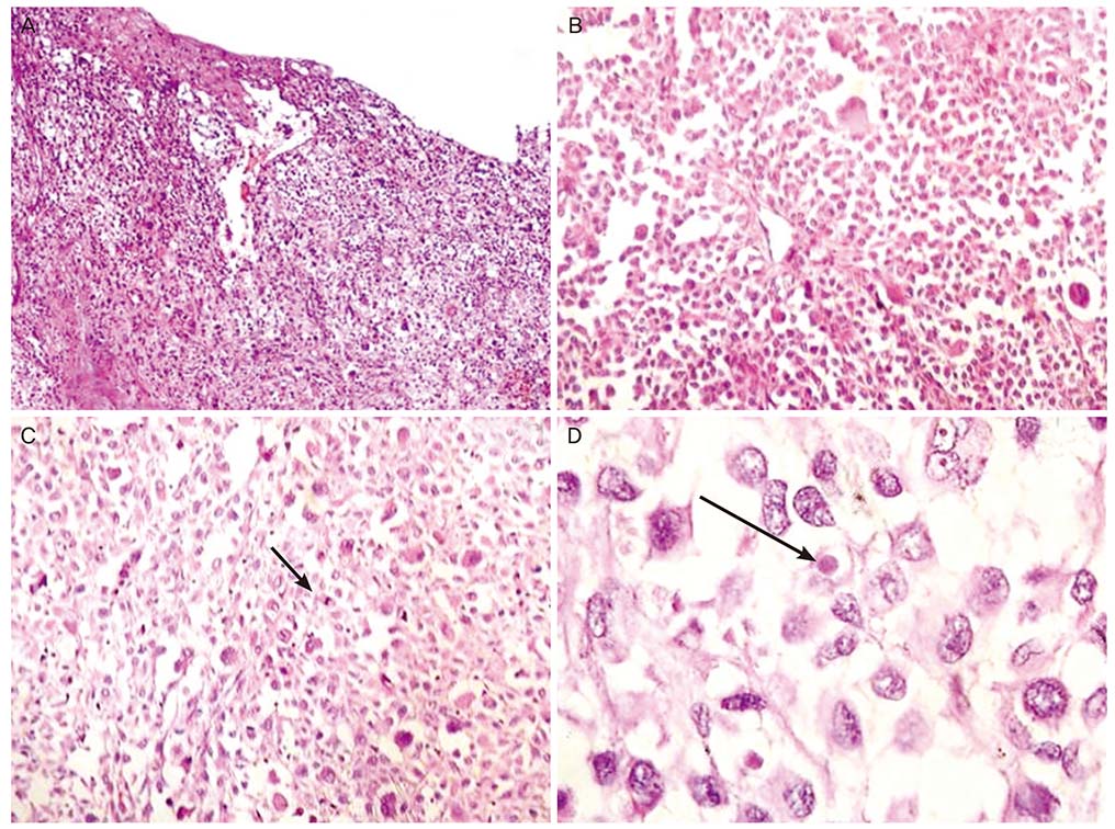
Fig. 1 Hematoxylin and eosin stained sections show (A) a tumor in the subepithelium with junctional activity and surface ulceration. (B) Tumor cells arranged in cords with intervening thin fibrous septa. Cells are plasmacytoid with eccentrically-placed nuclei and abundant cytoplasm. (C) Numerous mitotic figures (arrow). (D) Tumor cells have prominent eosinophilic nuclei and few hyaline globules (arrow).
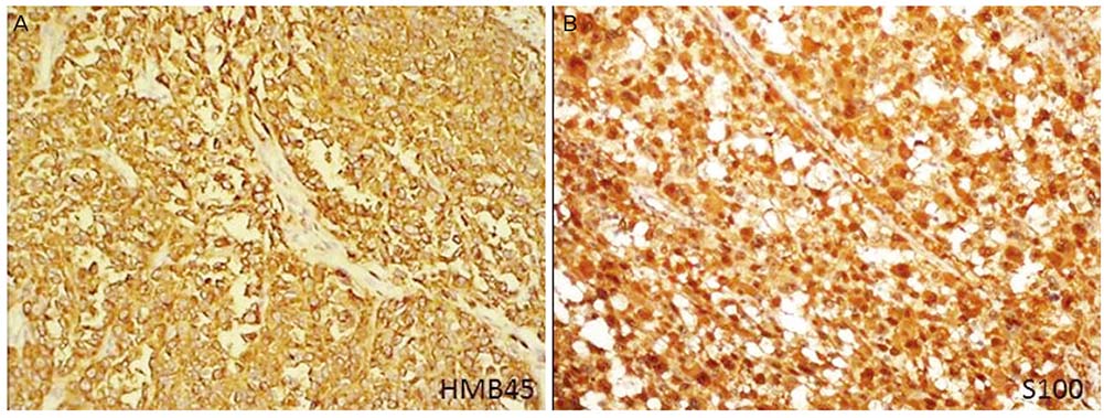
Fig. 2 Immunohistochemistry: (A) Tumor cell cytoplasm is positive for human melanoma black 45 (HMB45) and (B) nucleus and cytoplasm are positive for S-100
Reference
-
1. Mihajlovic M, Vlajkovic S, Jovanovic P, Stefanovic V. Primary mucosal melanomas: a comprehensive review. Int J Clin Exp Pathol. 2012; 5:739–753.2. Schmidt M, Honig A, Schwab M, Adam P, Dietl J. Primary vaginal melanoma: a case report and literature review. Eur J Gynaecol Oncol. 2008; 29:285–288.3. Samolis S, Panagopoulos P, Kanellopoulos N, Papastefanou I, Karadaglis S, Katsoulis M. Primary melanoma of the vagina: a case report. Eur J Gynaecol Oncol. 2010; 31:233–234.4. Kühn F, Dieterich M, Klar E, Gerber B, Prinz C. Primary malignant vaginal melanoma - case report and review of the literature. Geburtshilfe Frauenheilkd. 2012; 72:740–743.
Article5. Whiteman DC, Pavan WJ, Bastian BC. The melanomas: a synthesis of epidemiological, clinical, histopathological, genetic, and biological aspects, supporting distinct subtypes, causal pathways, and cells of origin. Pigment Cell Melanoma Res. 2011; 24:879–897.
Article6. Gökaslan H, Sişmanoğlu A, Pekin T, Kaya H, Ceyhan N. Primary malignant melanoma of the vagina: a case report and review of the current treatment options. Eur J Obstet Gynecol Reprod Biol. 2005; 121:243–248.
Article7. Sanchez AA, Wu TT, Prieto VG, Rashid A, Hamilton SR, Wang H. Comparison of primary and metastatic malignant melanoma of the esophagus: clinicopathologic review of 10 cases. Arch Pathol Lab Med. 2008; 132:1623–1629.
Article8. Allén AC, Spitz S. Malignant melanoma; a clinicopathological analysis of the criteria for diagnosis and prognosis. Cancer. 1953; 6:1–45.
Article9. Hodi FS, O'Day SJ, McDermott DF, Weber RW, Sosman JA, Haanen JB, et al. Improved survival with ipilimumab in patients with metastatic melanoma. N Engl J Med. 2010; 363:711–723.
Article
