Reconstructed Three-Dimensional Images and Parameters of Individual Erythrocytes Using Optical Diffraction Tomography Microscopy
- Affiliations
-
- 1Department of Laboratory Medicine, University of Ulsan College of Medicine and Asan Medical Center, Seoul, Korea. ssjang@amc.seoul.kr
- 2Department of Physics, Korea Advanced Institute of Science and Technology, Daejeon, Korea.
- KMID: 2425981
- DOI: http://doi.org/10.3343/alm.2019.39.2.223
Abstract
- No abstract available.
Figure
-
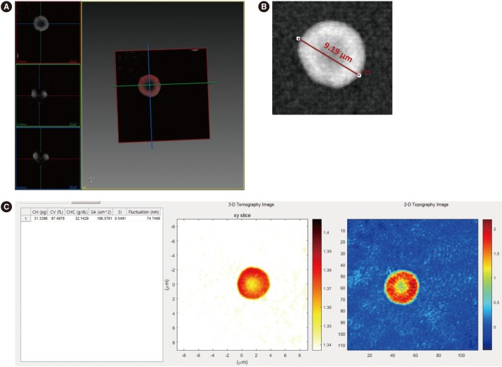
Fig. 1 Erythrocyte visualization and measurement of parameters from 3-D RI tomograms: (A) Measuring the diameter directly from the erythrocyte image. (B) Measuring various kinds of parameters calculated from 3-D and 2-D RI tomograms of an erythrocyte. (C) Output of the commercial software used (Tomostudio, Tomocube, Inc., Daejeon, Korea).Abbreviations: 3-D, three-dimensional; 2-D, two-dimensional; CH, corpuscular hemoglobin content; CV, corpuscular volume; CHC, corpuscular hemoglobin concentration; SA, surface area; SI, sphericity.
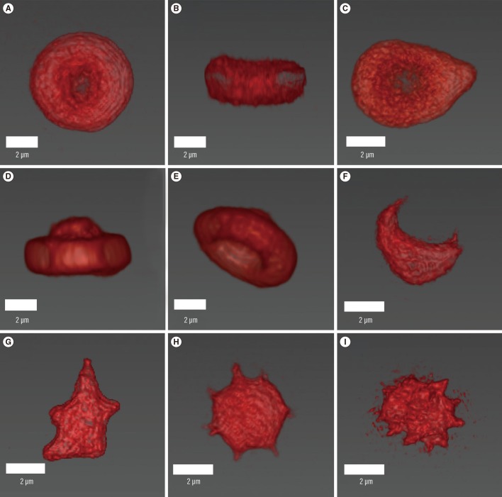
Fig. 2 Reconstructed three-dimensional refractive index tomograms of erythrocytes. Upper and side views of a normal erythrocyte (A, B), upper view of a teardrop cell (C), upper and side views of a target cell (D, E), side view of a schistocytes (F, G), an acanthocyte (H), and a burr cell (I). The reconstructed erythrocyte images were colored red by the authors' choice; they were originally greyscale images obtained using a commercial software (Tomostudio, Tomocube, Inc., Daejeon, Korea).
Reference
-
1. Salsbury AJ, Clarke JA. New method for detecting changes in the surface appearance of human red blood cells. J Clin Pathol. 1967; 20:603–610. PMID: 5628855.2. Goldstein JI, Newbury DE, Echlin P, Joy DC, Fiori C, Lifshin E. Scanning electron microscopy and X-ray analysis. A text for biologists, material scientists and geologists. 2nd ed. New York: Plenum Press;1992. p. 579–611.3. Park Y, Diez-Silva M, Fu D, Popescu G, Choi W, Barman I, et al. Static and dynamic light scattering of healthy and malaria-parasite invaded red blood cells. J Biomed Opt. 2015; 15:020506.4. Kim Y, Shim H, Kim K, Park H, Jang S, Park Y. Profiling individual human red blood cells using common-path diffraction optical tomography. Sci Rep. 2014; 4:6659. PMID: 25322756.5. Park H, Lee S, Ji M, Kim K, Son Y, Jang S, et al. Measuring cell surface area and deformability of individual human red blood cells over blood storage using quantitative phase imaging. Sci Rep. 2016; 6:34257. PMID: 27698484.6. Lee S, Park H, Kim K, Sohn Y, Jang S, Park Y. Refractive index tomograms and dynamic membrane fluctuations of red blood cells from patients with diabetes mellitus. Sci Rep. 2017; 7:1039. PMID: 28432323.
- Full Text Links
-
- Actions
-
Cited
- CITED
-
- Close
- Share
-
- Similar articles
-
- Unique Red Blood Cell Morphology Detected in a Patient with Myelodysplastic Syndrome by Three-dimensional Refractive Index Tomography
- Difference in glenoid retroversion between two-dimensional axial computed tomography and three-dimensional reconstructed images
- The Quality of Reconstructed 3D Images in Multidetector-Row Helical CT: Experimental Study Involving Scan Parameters
- Comparative study of glenoid version and inclination using two-dimensional images from computed tomography and three-dimensional reconstructed bone models
- Application of Three-dimensional Reconstruction in Esophageal Foreign Bodies
