The Findings of Ultrasonography, CT and Magnetic Resonance Imaging of Carotidynia: A Case Report
- Affiliations
-
- 1Department of Radiology, Hallym University College of Medicine, Chuncheon Sacred Heart Hospital, Chuncheon, Korea. khc@hallym.or.kr
Abstract
- Carotidynia is a rare disease representing various aspects of pain around the carotid artery bifurcation. The pain in the carotid bifurcation area is a nonspecific symptom, and various diseases causing similar symptoms in the corresponding area should be distinguished. Mostly, carotidynia is diagnosed by history taking and physical examination, but it is not easy to distinguish carotidynia from other diseases without imaging study. Therefore, there are quite a number of inadequate treatments due to a considerable number of misdiagnosis, as infectious diseases. In US, CT scans, and MRI examinations, the authors experienced a patient who showed the outer thickening wall of blood vessels around the carotid artery, near the carotid artery bifurcation, and was diagnosed as carotidynia with clinical findings. In the follow-up that was carried out two weeks later, the thickness of the lesions was significantly decreased. Imaging studies are helpful in the differential diagnosis because carotidynia shows relatively characteristic imaging findings.
MeSH Terms
Figure
-
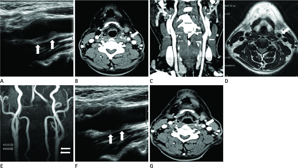
Fig. 1 A 49-year-old man with left neck pain with tenderness. A. Initial ultrasonography show hypo-echoic thickening of outer wall surrounding the left distal common carotid artery and more prominent thickening in medial perivascular area (arrows). Because ultrasonographic scanning is conducted from lateral to medial direction of neck, the arrows indicating area correspond to medial perivascular region of distal common carotid artery. B. Initial contrast-enhanced axial CT scan shows perivascular thickening with mild enhancement surrounding the left distal common carotid artery (arrow). C. Three-dimensional-reformated coronal image of initial contrast-enhanced CT shows mild luminal narrowing and perivascular thickening with mild enhancement surrounding the left distal common carotid artery and proximal internal carotid artery (arrows), compared to the opposite side (hollow arrows). D. Initial axial T2-weighted image shows well marginated perivascular hight signal intensity surrounding the left distal common carotid artery (arrow). E. On MRA image, there is mild luminal narrowing in affected artery (arrows), compared to the opposite side (hollow arrows). However, there is not evident as the CT findings. F, G. Follow-up ultrasonography (F) and CT (G) after 2 weeks show marked resolution of abnormal perivascular thickening (arrows).
Reference
-
1. Fay T. Atypical neuralgia. Arch Neurol Psychiatry. 1927; 18:309–315.2. Headache Classification Committee of the International Headache Society. Classification and diagnostic criteria for headache disorders, cranial neuralgias and facial pain. Cephalalgia. 1988; 8:Suppl 7. 1–96.3. Biousse V, Bousser MG. The myth of carotidynia. Neurology. 1994; 44:993–995.4. Upton PD, Smith JG, Charnock DR. Histologic confirmation of carotidynia. Otolaryngol Head Neck Surg. 2003; 129:443–444.5. Burton BS, Syms MJ, Petermann GW, Burgess LP. MR imaging of patients with carotidynia. AJNR Am J Neuroradiol. 2000; 21:766–769.6. Kuhn J, Harzheim A, Horz R, Bewermeyer H. MRI and ultrasonographic imaging of a patient with carotidynia. Cephalalgia. 2006; 26:483–485.7. Kosaka N, Sagoh T, Uematsu H, Kimura H, Miyayama S, Noguchi M, et al. Imaging by multiple modalities of patients with a carotidynia syndrome. Eur Radiol. 2007; 17:2430–2433.8. Pipitone N, Versari A, Salvarani C. Role of imaging studies in the diagnosis and follow-up of large-vessel vasculitis: an update. Rheumatology (Oxford). 2008; 47:403–408.9. Stanbro M, Gray BH, Kellicut DC. Carotidynia: revisiting an unfamiliar entity. Ann Vasc Surg. 2011; 25:1144–1153.10. Emmanuelli JL, Gutierrez JR, Chiossone JA, Chiossone E. Carotidynia: a frequently overlooked or misdiagnosed syndrome. Ear Nose Throat J. 1998; 77:462–464. 466469
- Full Text Links
-
- Actions
-
Cited
- CITED
-
- Close
- Share
-
- Similar articles
-
- Carotidynia presenting with acute ischemic stroke after carotid sinus massage
- Metastasis of Rhabdomyosarcoma to the Male Breast: a Case Report with Magnetic Resonance Imaging Findings
- Recurrent Primary Pleomorphic Liposarcoma of the Breast: A Case Report with Imaging Findings
- A study on the comparision of various imaging methods for the staging of renal cell carcinoma
- Castleman's Disease in the Sigmoid Mesocolon: A Case Report
