Image Quality and Radiation Dose of High-Pitch Dual-Source Spiral Cardiothoracic Computed Tomography in Young Children with Congenital Heart Disease: Comparison of Non-Electrocardiography Synchronization and Prospective Electrocardiography Triggering
- Affiliations
-
- 1Department of Radiology and Research Institute of Radiology, University of Ulsan College of Medicine, Asan Medical Center, Seoul 05505, Korea. ghw68@hanmail.net
- KMID: 2424843
- DOI: http://doi.org/10.3348/kjr.2018.19.6.1031
Abstract
OBJECTIVE
To compare image quality and radiation dose of high-pitch dual-source spiral cardiothoracic computed tomography (CT) between non-electrocardiography (ECG)-synchronized and prospectively ECG-triggered data acquisitions in young children with congenital heart disease.
MATERIALS AND METHODS
Eighty-six children (≤ 3 years) with congenital heart disease who underwent high-pitch dual-source spiral cardiothoracic CT were included in this retrospective study. They were divided into two groups (n = 43 for each; group 1 with non-ECG-synchronization and group 2 with prospective ECG triggering). Patient-related parameters, radiation dose, and image quality were compared between the two groups.
RESULTS
There were no significant differences in patient-related parameters including age, cross-sectional area, body density, and water-equivalent area between the two groups (p > 0.05). Regarding radiation dose parameters, only volume CT dose index values were significantly different between group 1 (1.13 ± 0.09 mGy) and group 2 (1.07 ± 0.12 mGy, p < 0.02). Among image quality parameters, significantly higher image noise (3.8 ± 0.7 Hounsfield units [HU] vs. 3.3 ± 0.6 HU, p < 0.001), significantly lower signal-to-noise ratio (105.0 ± 28.9 vs. 134.1 ± 44.4, p = 0.001) and contrast-to-noise ratio (84.5 ± 27.2 vs. 110.1 ± 43.2, p = 0.002), and significantly less diaphragm motion artifacts (3.8 ± 0.5 vs. 3.7 ± 0.4, p < 0.04) were found in group 1 compared with group 2. Image quality grades of cardiac structures, coronary arteries, ascending aorta, pulmonary trunk, lung markings, and chest wall showed no significant difference between groups (p > 0.05).
CONCLUSION
In high-pitch dual-source spiral pediatric cardiothoracic CT, additional ECG triggering does not substantially reduce motion artifacts in young children with congenital heart disease.
Keyword
MeSH Terms
Figure
-
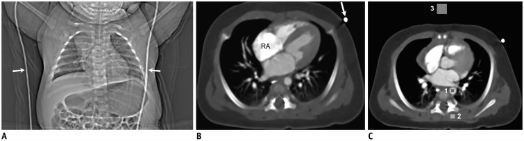
Fig. 1 77-day-old boy with coarctation of aorta.A. CT scout image shows ECG cables (arrows) for prospective ECG triggering. ECG electrodes placed on both arms are not shown on CT scout image. B. Axial CT image showing left-side ECG cable (arrow) causing mild streak artifact. Mild streak artifacts also are shown around RA. As result, degree of streak artifacts was assessed as grade 3 indicating mildly degraded image quality. C. Axial CT image at level of aortic sinus shows locations of three rectangular regions of interest for measuring CT densities in descending aorta (1), paraspinal muscle (2), and air (3). CT = computed tomography, ECG = electrocardiography, RA = right atrium
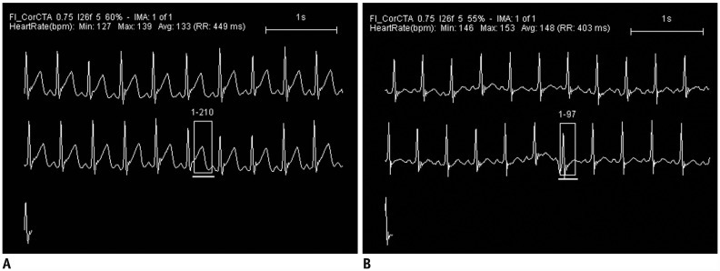
Fig. 2 Scan period positions in prospectively ECG-triggered high-pitch dual-source cardiothoracic CT scanning on ECG.A. In 77-day-old boy with coarctation of aorta and cervical aortic arch, scanning period on ECG, indicated by rectangle, is optimally located, starting from T wave and finishing with P wave peak. B. In contrast, scanning period on ECG, indicated by rectangle, is poorly positioned, overlapping with R wave in 13-day-old boy with hypoplastic left heart syndrome.
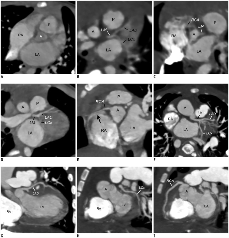
Fig. 3 Coronary artery motion artifact grading of high-pitch dual-source cardiothoracic CT.A. Oblique coronal CT image acquired with prospective ECG triggering in 35-day-old boy with coarctation of aorta demonstrates severe motion artifacts on coronary arteries that corresponded to grade 1. Oblique CT images (B, C) acquired without ECG synchronization in 1-day-old girl with coarctation of aorta illustrate moderate motion artifacts on coronary arteries, especially right coronary artery, which correspond to grade 2. Oblique CT images (D, E) acquired without ECG synchronization in an 11-month-old boy with surgically closed atrial septal defect reveal mild motion artifacts (grade 3), especially on right coronary artery, including doubling artifact (arrow) at proximal segment. Oblique CT images (F–I) acquired without ECG synchronization in 6-month-old boy with double-outlet right ventricle show no motion artifacts on coronary arteries including left main artery, left anterior descending artery, left circumflex artery, and right coronary artery that corresponded to grade 4. A = ascending aorta, LA = left atrium, LAD = left anterior descending artery, LCx = left circumflex artery, LM = left main artery, LV = left ventricle, P = pulmonary trunk, RCA = right coronary artery
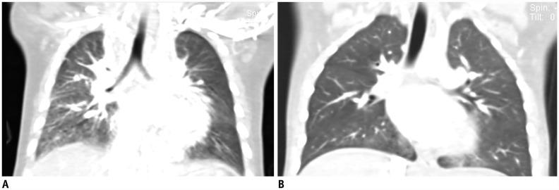
Fig. 4 Lung window setting CT images illustrating motion artifacts in lung markings and diaphragms.A. Coronal CT image acquired without ECG synchronization in 6-month-old boy with functional single ventricle shows moderate degrees of motion artifacts (grade 2) in lung markings as well as in diaphragm. B. Coronal CT image acquired without ECG synchronization in 3-year-old boy with repaired coarctation of aorta displays no motion artifacts (grade 4) in lung markings as well as diaphragm.
Cited by 1 articles
-
User-Friendly Vendor-Specific Guideline for Pediatric Cardiothoracic Computed Tomography Provided by the Asian Society of Cardiovascular Imaging Congenital Heart Disease Study Group: Part 1. Imaging Techniques
Sun Hwa Hong, Hyun Woo Goo, Eriko Maeda, Ki Seok Choo, I-Chen Tsai,
Korean J Radiol. 2019;20(2):190-204. doi: 10.3348/kjr.2018.0571.
Reference
-
1. Goo HW, Park IS, Ko JK, Kim YH, Seo DM, Yun TJ, et al. CT of congenital heart disease: normal anatomy and typical pathologic conditions. Radiographics. 2003; 23 Spec No:S147–S165. PMID: 14557509.2. Goo HW. State-of-the-art CT imaging techniques for congenital heart disease. Korean J Radiol. 2010; 11:4–18. PMID: 20046490.3. Goo HW, Yang DH. Coronary artery visibility in free-breathing young children with congenital heart disease on cardiac 64-slice CT: dual-source ECG-triggered sequential scan vs. single-source non-ECG-synchronized spiral scan. Pediatr Radiol. 2010; 40:1670–1680. PMID: 20464385.4. Goo HW. Current trends in cardiac CT in children. Acta Radiol. 2013; 54:1055–1062. PMID: 23104372.
Article5. Goo HW. Coronary artery imaging in children. Korean J Radiol. 2015; 16:239–250. PMID: 25741188.
Article6. Bang JH, Park JJ, Goo HW. Evaluation of commissural malalignment of aortic-pulmonary sinus using cardiac CT for arterial switch operation: comparison with transthoracic echocardiography. Pediatr Radiol. 2017; 47:556–564. PMID: 28243677.7. Goo HW. Identification of coronary artery anatomy on dual-source cardiac computed tomography before arterial switch operation in newborns and young infants: comparison with transthoracic echocardiography. Pediatr Radiol. 2018; 48:176–185. PMID: 29032431.8. Flohr TG, Leng S, Yu L, Aiimendinger T, Bruder H, Petersilka M, et al. Dual-source spiral CT with pitch up to 3.2 and 75 ms temporal resolution: image reconstruction and assessment of image quality. Med Phys. 2009; 36:5641–5653. PMID: 20095277.9. Goetti R, Feuchtner G, Stolzmann P, Desbiolles L, Fischer MA, Karlo C, et al. High-pitch dual-source CT coronary angiography: systolic data acquisition at high heart rates. Eur Radiol. 2010; 20:2565–2571. PMID: 20585785.10. Scharf M, Bink R, May MS, Hentschke C, Achenbach S, Uder M, et al. High-pitch thoracic CT with simultaneous assessment of coronary arteries: effect of heart rate and heart rate variability on image quality and diagnostic accuracy. JACC Cardiovasc Imaging. 2011; 4:602–609. PMID: 21679894.11. Bamberg F, Marcus R, Sommer W, Schwarz F, Nikolaou K, Becker CR, et al. Diagnostic image quality of a comprehensive high-pitch dual-spiral cardiothoracic CT protocol in patients with undifferentiated acute chest pain. Eur J Radiol. 2012; 81:3697–3702. PMID: 21196093.12. Kröpil P, Rojas CA, Ghoshhajra B, Lanzman RS, Miese FR, Scherer A, et al. Prospectively ECG-triggered high-pitch spiral acquisition for cardiac CT angiography in routine clinical practice: initial results. J Thorac Imaging. 2012; 27:194–201. PMID: 21964497.13. Sun K, Han RJ, Ma LJ, Wang LJ, Li LG, Chen JH. Prospectively electrocardiogram-gated high-pitch spiral acquisition mode dual-source CT coronary angiography in patients with high heart rates: comparison with retrospective electrocardiogram-gated spiral acquisition mode. Korean J Radiol. 2012; 13:684–693. PMID: 23118566.14. Bischoff B, Meinel FG, Del Prete A, Reiser MF, Becker HC. High-pitch coronary CT angiography in dual-source CT during free breathing vs. breath holding in patients with low heart rates. Eur J Radiol. 2013; 82:2217–2221. PMID: 24075783.15. Wang Q, Qin J, He B, Zhou Y, Yang JJ, Hou XL, et al. Double prospectively ECG-triggered high-pitch spiral acquisition for CT coronary angiography: initial experience. Clin Radiol. 2013; 68:792–798. PMID: 23601956.16. St Noble V, Douraghi-Zadeh D, Padley SP, Rubens MB, Nicol ED. Maximizing the clinical benefit of high-pitch, single-heartbeat CT coronary angiography in clinical practice. Clin Radiol. 2014; 69:674–677. PMID: 24581960.17. Deseive S, Pugliese F, Meave A, Alexanderson E, Martinoff S, Hadamitzky M, et al. Image quality and radiation dose of a prospectively electrocardiography-triggered high-pitch data acquisition strategy for coronary CT angiography: the multicenter, randomized PROTECTION IV study. J Cardiovasc Comput Tomogr. 2015; 9:278–285. PMID: 25926015.18. Lell MM, May M, Deak P, Alibek S, Kuefner M, Kuettner A, et al. High-pitch spiral computed tomography: effect on image quality and radiation dose in pediatric chest computed tomography. Invest Radiol. 2011; 46:116–123. PMID: 20856124.19. Nie P, Wang X, Cheng Z, Ji X, Duan Y, Chen J. Accuracy, image quality and radiation dose comparison of high-pitch spiral and sequential acquisition on 128-slice dual-source CT angiography in children with congenital heart disease. Eur Radiol. 2012; 22:2057–2066. PMID: 22592808.20. Zheng M, Zhao H, Xu J, Wu Y, Li J. Image quality of ultra-low-dose dual-source CT angiography using high-pitch spiral acquisition and iterative reconstruction in young children with congenital heart disease. J Cardiovasc Comput Tomogr. 2013; 7:376–382. PMID: 24331933.21. Xu J, Zhao H, Wang X, Bai Y, Liu L, Liu Y, et al. Accuracy, image quality, and radiation dose of prospectively ECG-triggered high-pitch dual-source CT angiography in infants and children with complex coarctation of the aorta. Acad Radiol. 2014; 21:1248–1254. PMID: 25097011.22. Nie P, Yang G, Wang X, Duan Y, Xu W, Li H, et al. Application of prospective ECG-gated high-pitch 128-slice dual-source CT angiography in the diagnosis of congenital extracardiac vascular anomalies in infants and children. PLoS One. 2014; 9:e115793. PMID: 25546178.23. Sriharan M, Lazoura O, Pavitt CW, Castellano I, Owens CM, Rubens MB, et al. Evaluation of high-pitch ungated pediatric cardiovascular computed tomography for the assessment of cardiac structures in neonates. J Thorac Imaging. 2016; 31:177–182. PMID: 27007667.24. Kanie Y, Sato S, Tada A, Kanazawa S. Image quality of coronary arteries on non-electrocardiography-gated high-pitch dual-source computed tomography in children with congenital heart disease. Pediatr Cardiol. 2017; 38:1393–1399. PMID: 28689328.25. Goo HW. Individualized volume CT dose index determined by cross-sectional area and mean density of the body to achieve uniform image noise of contrast-enhanced pediatric chest CT obtained at variable kV levels and with combined tube current modulation. Pediatr Radiol. 2011; 41:839–847. PMID: 21656275.26. Hui PKT, Goo HW, Du J, Ip JJK, Kanzaki S, Kim YJ, et al. Asian consortium on radiation dose of pediatric cardiac CT (ASCI-REDCARD). Pediatr Radiol. 2017; 47:899–910. PMID: 28435986.27. Goo HW. CT radiation dose optimization and estimation: an update for radiologists. Korean J Radiol. 2012; 13:1–11. PMID: 22247630.28. Beeres M, Wichmann JL, Frellesen C, Bucher AM, Albrecht M, Scholtz JE, et al. ECG-gated versus non-ECG-gated high-pitch dual-source CT for whole body CT angiography (CTA). Acad Radiol. 2016; 23:163–167. PMID: 26548854.29. Wielandner A, Beitzke D, Schernthaner R, Wolf F, Langenberger C, Stadler A, et al. Is ECG triggering for motion artefact reduction in dual-source CT angiography of the ascending aorta still required with high-pitch scanning? The role of ECG-gating in high-pitch dual-source CT of the ascending aorta. Br J Radiol. 2016; 6. 27. [Epub ahead of print]. DOI: 10.1259/bjr.20160174.30. Farshad-Amacker NA, Alkadhi H, Leschka S, Frauenfelder T. Effect of high-pitch dual-source CT to compensate motion artifacts: a phantom study. Acad Radiol. 2013; 20:1234–1239. PMID: 24029055.31. Koch K, Oellig F, Oberholzer K, Bender P, Kunz P, Mildenberger P, et al. Assessment of right ventricular function by 16-detector-row CT: comparison with magnetic resonance imaging. Eur Radiol. 2005; 15:312–318. PMID: 15565315.32. Goo HW, Park SH. Semiautomatic three-dimensional CT ventricular volumetry in patients with congenital heart disease: agreement between two methods with different user interaction. Int J Cardiovasc Imaging. 2015; 31(Suppl 2):223–232. PMID: 26319216.33. Goo HW, Allmendinger T. Combined electrocardiography- and respiratory-triggered CT of the lung to reduce respiratory misregistration artifacts between imaging slabs in free-breathing children: initial experience. Korean J Radiol. 2017; 18:860–866. PMID: 28860904.34. Beeres M, Loch M, Schulz B, Kerl M, Al-Butmeh F, Bodelle B, et al. Bolus timing in high-pitch CT angiography of the aorta. Eur J Radiol. 2013; 82:1028–1033. PMID: 23375815.
Article35. Beeres M, Williams K, Bauer RW, Scholtz J, Kaup M, Gruber-Rouh T, et al. First clinical evaluation of high-pitch dual-source computed tomographic angiography comparing automated tube potential selection with automated tube current modulation. J Comput Assist Tomogr. 2015; 39:624–628. PMID: 25955395.
- Full Text Links
-
- Actions
-
Cited
- CITED
-
- Close
- Share
-
- Similar articles
-
- Prospectively Electrocardiogram-Gated High-Pitch Spiral Acquisition Mode Dual-Source CT Coronary Angiography in Patients with High Heart Rates: Comparison with Retrospective Electrocardiogram-Gated Spiral Acquisition Mode
- Comparison of Chest Pain Protocols for Electrocardiography-Gated Dual-Source Cardiothoracic CT in Children and Adults: The Effect of Tube Current Saturation on Radiation Dose Reduction
- Combined Electrocardiography- and Respiratory-Triggered CT of the Lung to Reduce Respiratory Misregistration Artifacts between Imaging Slabs in Free-Breathing Children: Initial Experience
- Image Quality and Radiation Dose of Lower Extremity CT Angiography in 128 Slice Dual-Source CT: Comparison of High Pitch and Low Pitch
- State-of-the-Art CT Imaging Techniques for Congenital Heart Disease
