J Korean Fract Soc.
2017 Jul;30(3):142-145. 10.12671/jkfs.2017.30.3.142.
Extensive Multiple Morel-Lavallée Lesions: A Case Report
- Affiliations
-
- 1Department of Orthopaedic Surgery, Guri Hospital, Hanyang University College of Medicine, Guri, Korea. hyparkys@hanyang.ac.kr
- KMID: 2424123
- DOI: http://doi.org/10.12671/jkfs.2017.30.3.142
Abstract
- Morel-Lavallée is a rare lesion caused by post-traumatic soft tissue injury. It usually occurs around the greater trochanter, and it occurs very rarely in the lumbar region. It is often difficult to be diagnosed in the emergency room. Delayed diagnosis may result in the need for open surgery. The authors report a patient with extensive multiple Morel-Lavallée lesions in the thoracolumbar, buttock, and thigh after trauma and provide a literature review.
Keyword
MeSH Terms
Figure
-

Fig. 1 Sagittal T2-weighted fat-suppressed image of the thoracolumbar spine (A) shows a large fluid collection within the subcutaneous tissues, from the T10 level to the S1 level. Coronal T2-weighted fat-suppressed image of the left thigh (B) and axial image of the right hip (C) show a fluid collection between the fascia and the subcutaneous layer.

Fig. 2 Intraoperative appearance of Morel-Lavallée lesions at the thoracolumbar spine (A) and left thigh (B).
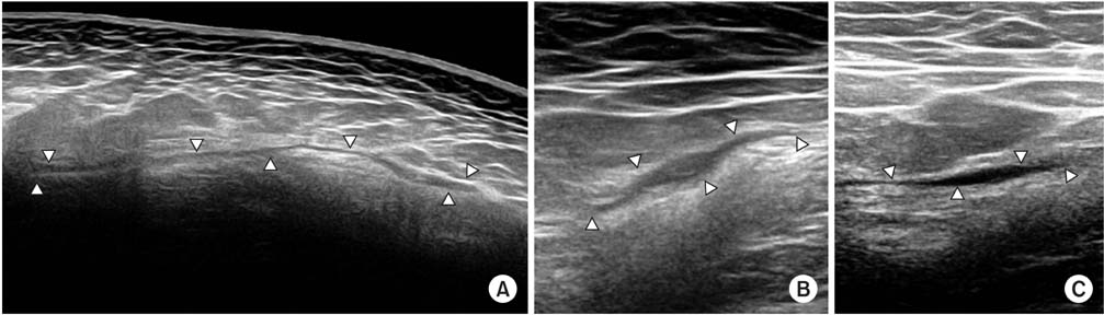
Fig. 3 Longitudinal ultrasound image of the thoracolumbar spine (A), and transverse ultrasound image of the left thigh (B) and right hip (C).
Reference
-
1. Letournel E, Judet R. Fractures of the acetabulum. 2nd ed. Berlin: Springer;1993.2. Parra JA, Fernandez MA, Encinas B, Rico M. Morel-Lavallée effusions in the thigh. Skeletal Radiol. 1997; 26:239–241.
Article3. Kalaci A, Karazincir S, Yanat AN. Long-standing Morel-Lavallée lesion of the thigh simulating a neoplasm. Clin Imaging. 2007; 31:287–291.
Article4. El Kininy W, Davy S, Sayana M. Unusual Morel-Lavallee lesion of the knee region in an elderly patient. BMJ Case Rep. 2017; 2017:bcr2016218577.
Article5. Luria S, Applbaum Y, Weil Y, Liebergall M, Peyser A. Talc sclerodhesis of persistent Morel-Lavallée lesions (posttraumatic pseudocysts): case report of 4 patients. J Orthop Trauma. 2006; 20:435–438.
Article6. Tseng S, Tornetta P 3rd. Percutaneous management of Morel-Lavallee lesions. J Bone Joint Surg Am. 2006; 88:92–96.
Article7. Hudson DA, Knottenbelt JD, Krige JE. Closed degloving injuries: results following conservative surgery. Plast Reconstr Surg. 1992; 89:853–855.8. Zairi F, Wang Z, Shedid D, Boubez G, Sunna T. Lumbar Morel-Lavallée lesion: case report and review of the literature. Orthop Traumatol Surg Res. 2016; 102:525–527.
Article9. Tran W, Foran J, Wang M, Schwartz A. Postsurgical bleeding following treatment of a chronic Morel-Lavallée lesion. Orthopedics. 2008; 31:814.10. Neal C, Jacobson JA, Brandon C, Klaume-Brigido M, Morag Y, Girish G. Sonography of Morel-Lavallée lesions. J Ultrasound Med. 2008; 27:1077–1081.
Article
- Full Text Links
-
- Actions
-
Cited
- CITED
-
- Close
- Share
-
- Similar articles
-
- Morel-Lavallée Lesion in the Sacrococcygeal Area with Associated Coccygeal Fracture
- Occurrence and Surgical Treatment of Massive Morel-Lavallée Lesion after Large-Volume Liposuction: A Rare Clinical Case
- Sclerotherapy Using Abnobaviscum for the Extensive Recurrent Chronic Morel-Lavallée Lesions - A Case Report -
- Posttraumatic bilateral thigh Morel-Lavallée lesions without an underlying bone fracture in the United Kingdom: a case report
- Sonographic Findings of Morel-Lavallee Lesions
