Primary Neuroendocrine Tumor of the Extrahepatic Bile Duct
- Affiliations
-
- 1Department of Internal Medicine, Inje University Ilsan Paik Hospital, Goyang, Korea. lys0326@paik.ac.kr
- 2Department of Surgery, Inje University Ilsan Paik Hospital, Goyang, Korea.
- 3Department of Pathology, Inje University Ilsan Paik Hospital, Goyang, Korea.
- KMID: 2424116
- DOI: http://doi.org/10.4166/kjg.2018.72.4.222
Abstract
- No abstract available.
Figure
-
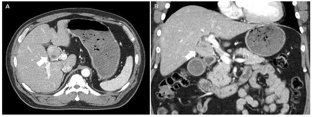
Fig. 1 (A, B) Contrast-enhanced CT image shows a 3.6×4.3 cm sized enhancing mass (arrows) in porta hepatis. CT, computed tomography.

Fig. 2 Magnetic resonance cholangiopancreatography shows low signal intensity mass involving the common hepatic duct (white arrow).
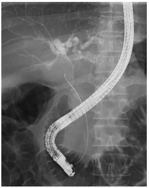
Fig. 3 Endoscopic retrograde cholangiogram reveals biliary stricture of the common hepatic duct.

Fig. 4 Gross finding of the resected tumor shows a 4.7×4.5×3.5 cm sized protruding mass into the bile duct.

Fig. 5 Histopathologic findings. (A) Tumor cells shows trabecular arrangement. Cytoplasms are plump and deeply eosinophilic. Nuceli have round, smooth outline and dark chromatin (H&E, ×200). (B) Tumor cell cytoplasms are strongly stained with synaptophysin immunohistochemistry, which indicates neuroendocrine differentiation (synaptophysin, ×200).
Reference
-
1. Carriaga MT, Henson DE. Liver, gallbladder, extrahepatic bile ducts, and pancreas. Cancer. 1995; 75:1 Suppl. 171–190.
Article2. Modlin IM, Sandor A. An analysis of 8305 cases of carcinoid tumors. Cancer. 1997; 79:813–829.
Article3. Michalopoulos N, Papavramidis TS, Karayannopoulou G, Pliakos I, Papavramidis ST, Kanellos I. Neuroendocrine tumors of extrahepatic biliary tract. Pathol Oncol Res. 2014; 20:765–775.
Article4. Lee SJ, Lee YS, Lee MG, Lee SH, Shin E, Hwang JH. Triple-tissue sampling during endoscopic retrograde cholangiopancreatography increases the overall diagnostic sensitivity for cholangiocarcinoma. Gut Liver. 2014; 8:669–673.
Article5. Abe T, Nirei A, Suzuki N, et al. Neuroendocrine tumor of the extrahepatic bile duct: a case report. Int J Surg Case Rep. 2017; 40:6–9.
Article6. Hubert C, Sempoux C, Berquin A, Deprez P, Jamar F, Gigot JF. Bile duct carcinoid tumors: an uncommon disease but with a good prognosis? Hepatogastroenterology. 2005; 52:1042–1047.7. Malecki EA, Acosta R, Twaddell W, Heller T, Manning MA, Darwin P. Endoscopic diagnosis of a biliary neuroendocrine tumor. Gastrointest Endosc. 2009; 70:1275–1276.
Article8. CRindi GAR, Bosman FT, Capella C, et al. Nomenclature and classification of neuroendocrine neoplasms of the digestive system. In : Bosman FT, Carneiro F, Hruban RH, Theise ND, editors. WHO Classification of Tumors of the Digestive System. 4th ed. Lyon: International Agency for Research on Cancer;2010. p. 13–14.9. Pawlik TM, Shah S, Eckhauser FE. Carcinoid tumor of the biliary tract: treating a rare cause of bile duct obstruction. Am Surg. 2003; 69:98–101.10. Khuroo S, Rashid A, Bali RS, Mushtaque M, Khuroo F. Carcinoid klatskin tumour: a rare cause of obstructive jaundice. Australas Med J. 2014; 7:243–246.
Article11. Chamberlain RS, Blumgart LH. Carcinoid tumors of the extrahepatic bile duct. A rare cause of malignant biliary obstruction. Cancer. 1999; 86:1959–1965.12. Lee KJ, Cho JH, Lee SH, et al. Clinicopathological characteristics of biliary neuroendocrine neoplasms: a multicenter study. Scand J Gastroenterol. 2017; 52:437–441.
Article
- Full Text Links
-
- Actions
-
Cited
- CITED
-
- Close
- Share
-
- Similar articles
-
- Extrahepatic Bile Duct Duplication with Intraductal Papillary Neoplasm: A Case Report
- Concurrent Yellow-to-white and Black Extrahepatic Bile Duct Stones
- Small-cell Neuroendocrine Carcinoma of the Extrahepatic Bile Duct: A Rare Case Report
- A case of extrahepatic bile duct hepatocellular carcinoma with no detectable primary hepatic tumor
- A Case of Multiple Papillary Adenocarcinoma of the Extrahepatic Bile Duct : Findings of ERCP
