J Korean Orthop Assoc.
2018 Oct;53(5):407-414. 10.4055/jkoa.2018.53.5.407.
Evaluation of the Outcomes according to Etiology in the Pediatric Pes Planovalgus after Lateral Column Lengthening: By Radiologic and Pedobarographic Measurements
- Affiliations
-
- 1Department of Orthopaedic Surgery, Chonnam National University Medical School, Gwangju, Korea. stjung@chonnam.ac.kr
- KMID: 2424099
- DOI: http://doi.org/10.4055/jkoa.2018.53.5.407
Abstract
- PURPOSE
Pes planovalgus is one of the most common foot deformities among pediatric orthopedic diseases and is divided into idiopathic and neuromuscular planovalgus according to its etiology. This study evaluated the radiologic and pedobarographic outcomes of the treatment for pes planovalgus in children treated with lateral column lengthening, compared the outcomes according to the etiology, and investigated the correlation between the radiologic and pedobarographic findings.
MATERIALS AND METHODS
Sixty-three patients (97 feet), who underwent lateral column lengthening, were divided into groups of 30 patients (47 feet) with neuromuscular disease and 33 patients (50 feet) with idiopathic etiology. The preoperative, immediately postoperative, 1 year, and 3 year follow-up radiographic measurements on the plain radiograph antero-posterior (AP) and lateral view were compared. In pedobarography, the foot pressures were subdivided into 4 areas to measure the contact time, contact area, peak pressure, and maximum force. The pre- and postoperative pedobarographic measurements were compared and the correlations between the radiographic and pedobarographic measurements were evaluated.
RESULTS
The radiographic index at the 1st postoperative year and 3rd postoperative follow-up did not show significant differences according to the etiology. In pedobarography, idiopathic planovalgus showed a significant increase in the maximum force in the hindfoot and forefoot. The correlation between the radiologic findings and pedobarographic findings was statistically significant between the tibiocalcaneal angle in the lateral view and the maximum force, and the contact area of hindfoot on pedobarography, between tibiocalcaneal angle in the lateral view and the contact area of the toes in idiopathic planovalgus. In neuromuscular planovalgus, the peak pressure in the hindfoot had a strong negative correlation with talonavicular coverage angle in the AP view and talo-1st metatarsal angle, and the talohorizontal angle in the lateral view.
CONCLUSION
Lateral column lengthening is an effective surgical procedure for flatfoot patients. On the other hand, the radiographic examination has limitations for accurate assessments of the postoperative results and prognosis. Qualitative and quantitative evaluations are available by pedobarography and it is a useful instrument for an evaluation of planovalgus when used in conjunction with radiography.
MeSH Terms
Figure
-
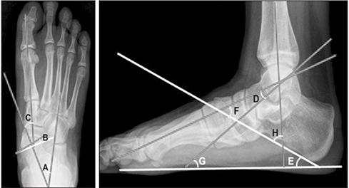
Figure 1 Radiographic parameters for an evaluation of pes planovalgus. A, talocalcaneal angle; B, talonavicular coverage angle; C, talo-1st metatarsal angle (antero-posterior view); D, talo-1st metatarsal angle (lateral view); E, calcaneal pitch; F, talocalcaneal angle; G, talo-horizontal angle; H, tibiocalcaneal angle.
Reference
-
1. Ananthakrisnan D, Ching R, Tencer A, Hansen ST Jr, Sangeorzan BJ. Subluxation of the talocalcaneal joint in adults who have symptomatic flatfoot. J Bone Joint Surg Am. 1999; 81:1147–1154.2. Andreacchio A, Orellana CA, Miller F, Bowen TR. Lateral column lengthening as treatment for planovalgus foot deformity in ambulatory children with spastic cerebral palsy. J Pediatr Orthop. 2000; 20:501–505.
Article3. Barry RJ, Scranton PE Jr. Flat feet in children. Clin Orthop Relat Res. 1983; (181):68–75.
Article4. Evans D. Calcaneo-valgus deformity. J Bone Joint Surg Br. 1975; 57:270–278.
Article5. Mosca VS. Calcaneal lengthening for valgus deformity of the hindfoot. Results in children who had severe, symptomatic flatfoot and skewfoot. J Bone Joint Surg Am. 1995; 77:500–512.
Article6. Sullivan JA. The child's foot. In : Morrissy RT, Weinstein SL, editors. Lovell and winter's pediatric orthopaedics. 4th ed. Philadelphia: Lippincott-Raven;1996. p. 1077–1136.7. Phillips GE. A review of elongation of os calcis for flat feet. J Bone Joint Surg Br. 1983; 65:15–18.
Article8. Frances JM, Feldman DS. Management of idiopathic and nonidiopathic flatfoot. Instr Course Lect. 2015; 64:429–440.9. Richter M, Zech S. Lengthening osteotomy of the calcaneus and flexor digitorum longus tendon transfer in flexible flatfoot deformity improves talo-1st metatarsal-index, clinical outcome and pedographic parameter. Foot Ankle Surg. 2013; 19:56–61.
Article10. Park ES, Kim HW, Park CI, Rha DW, Park CW. Dynamic foot pressure measurements for assessing foot deformity in persons with spastic cerebral palsy. Arch Phys Med Rehabil. 2006; 87:703–709.
Article11. Alvarez C, De Vera M, Chhina H, Black A. Normative data for the dynamic pedobarographic profiles of children. Gait Posture. 2008; 28:309–315.
Article12. Han SH, Jung M, Lee JW. Pedobarographic analysis in functional foot orthosis. J Korean Foot Ankle Soc. 2006; 10:125–132.13. Seol YJ, Jung ST, Yang HK, et al. Diagnostic availability of pedobarography and correlation of radiographic and pedobarographic measurements in pediatric flexible flatfoot. J Korean Orthop Assoc. 2014; 49:366–373.
Article
- Full Text Links
-
- Actions
-
Cited
- CITED
-
- Close
- Share
-
- Similar articles
-
- Surgical Treatments and Clinical Outcomes for Idiopathic Osteoarthritis of the Tarsometatarsal Joints
- Radiologic Measurement of Pes Cavus
- Surgical Treatment for Planovalgus Foot in Children with Generalized Ligamentous Laxity
- The Results of Treatment of Planovalgus Deformity by Modified Grice-Green Procedure
- Revision Surgery for Recurrent Pain after Excision of the Accessory Navicular and Relocation of the Tibialis Posterior Tendon
